| Muscle | Origin | Insertion | Innervation | Action |
|---|---|---|---|---|
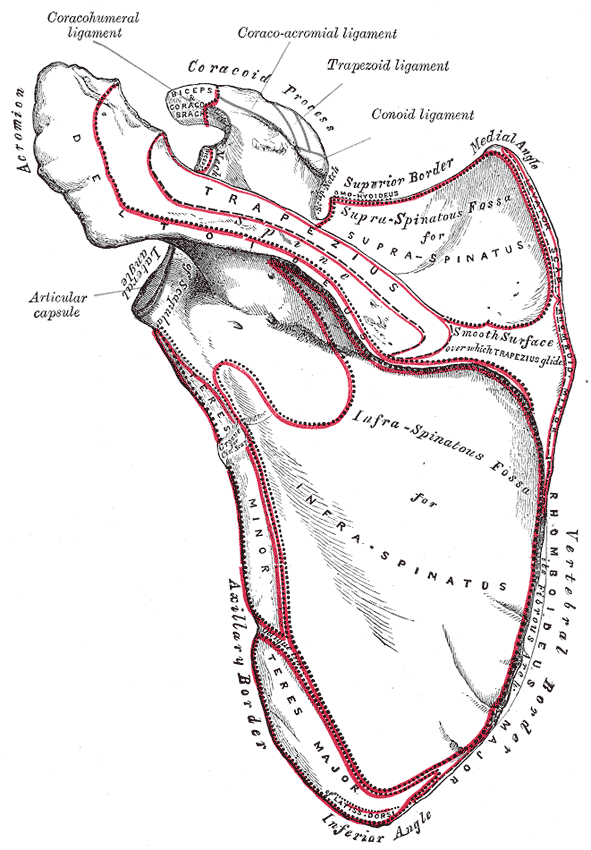
| Infraspinatus | Infraspinous Fossa of Scapula | Greater tubercle of Humerus | Suprascapular n. C4 - C6 |
GHJ: ER, Stabilization |
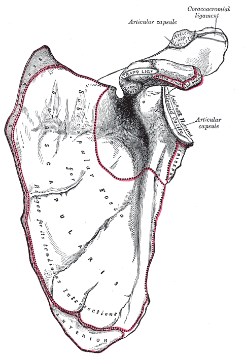
| Subscapularis | Subscapular fossa | Lesser tubercle of Humerus | Upper Subscapular n. Lower Subscapular n. C5 - C6 |
GHJ: IR, Stabilization |
| Supraspinatus | Supraspinous Fossa of scapula | Greater tubercle of Humerus | Suprascapular n. C4 - C6 |
GHJ: Initiates abduction, Stabilization |
| Teres minor | Greater tubercle of Humerus | Axillary n. C5 - C6 |
GHJ: ER, Weak adduction |
Shoulder MSK
Musculoskeletal overview
There are 3 major axes of the shoulder:
- Transverse Axis: Allows movements of flexion and extension in the sagittal plane7
- Antero-posterior axis: Allows movements in the frontal plane7
- Vertical axis: runs through the intersection of the sagittal and frontal planes and controls the movements of flexion and extension7.
Bones
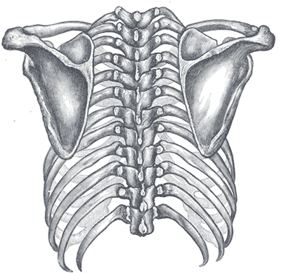
The Shoulder complex is made up of 3 bones:
Humerus
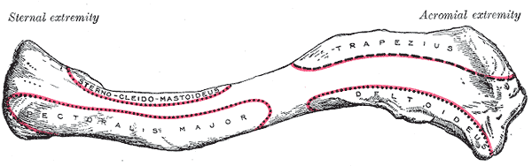
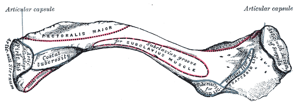
Clavicle
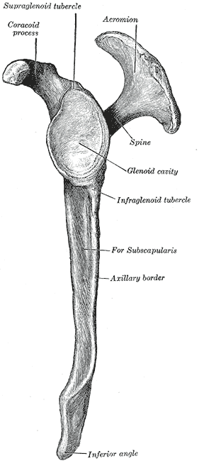
Scapula
Joints
The Shoulder complex is made up of 3 joints and 1 pseudo joint:
- SCJ provides gross movement of the clavicle8.
- The ACJ provides fine motor control of the clavicle8.
- The glenohumeral joint
- The scapulothoracic (pseudo) joint
. Anterior aspect.gif)
Sternoclavicular Joint
Acromioclavicular joint
The acromioclavicular joint (ACJ) is an articulation
The ACJ has poor stability since the joint surfaces do not interlock making it prone to dislocations7.
The acromion aspect of the joint is slightly convex and faces supero-antero-medially7. The clavicular aspect is flat or slightly convex and faces infero-postero-laterally
Since these two facets can be flat or even both convex, they have poor joint congruity and in 30% of cases, an intra-articular fibrocartilage meniscus is present to improve congruency7.
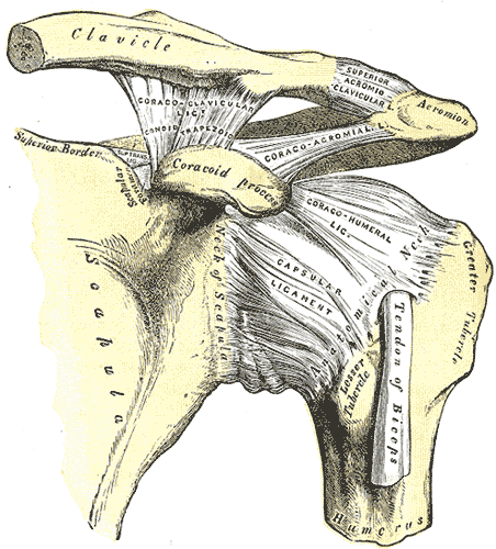
This joint’s stability is augmented by 2 extra-articular ligaments for stability: trapezoid lig. and conoid lig.7.
Glenohumeral joint
Scapulothoraic Joint
The Scapulothoracic Joint is considered a “pseudo joint”2
Physiologic Area
- Suprahumeral/Subacromial space
Ligaments
“(GH Ligs) Together with the coracohumeral ligament, these ligaments form a Z-pattern on the anterior aspect of the shoulder (Fig. 16-6).3 In the midrange of rotation, the G-H ligaments are relatively lax”2
Flexion & Extension
Flexion and extension are movements of the shoulder that occur in the sagittal plane about a transverse axis7.
| Movement | GHJ | Scapulohumeral | Total |
|---|---|---|---|
| Extension | 45-50° | ||
| Flexion | 120° | 60° | 180° |
Abduction & Adduction
Abduction and adduction of the shoulder refers to frontal/coronal plane movement occurring about an antero-posterior axis.
Normal abduction is 180°7.
During abduction, the should complex made up of the GHJ, ACJ and STJ work together in 3 distinct phases to complete the whole range of motion.
- 0-60° abduction takes place only at the glenohumeral joint7.
- 60-180° is a combination of glenohumeral joint and the scapulothoracic joint7.
Abduction
There are 4 abductor muscles that form a force coupling of shoulder abduction7.
- Glenohumeral
- deltoid
- Supraspinatus
- Scapulothoracic joint (upward rotation)
- Serratus anterior
- Upper trapezius
During abduction, the subscapularis, infraspinatus, and teres minor draw the humeral the infero-medially during abduction7.
Note
The biceps brachii long head is also involved in abduction and a rupture will result in ~20% decrease in abduction strength7.
Pure abduction
Pure abduction occurs in the frontal plane.
Scapular plane abduction
Scapular plane exists between the sagittal and frontal planes. 30° from the frontal plane7.
External rotation into abduction
- II (anterior deltoid) contracts at the start
Note
IV and V (Posterior deltoid) do not contract at all during this movement7.
IR to abduction
“recruitment order reverses”7.
Axial Rotation
The “neutral” position in axial rotation is where the elbow is flexed to 90° and the forearm is in the sagittal plane.
Note
Another “neutral” position is at 30° of internal rotation where the rotator muscles are at equilibrium7.
External rotation
Horizontal abduction & adduction
Stabilization
Passive
The shoulder maintains its passive stability by using multiple structures arranged reciprocally so when one structure is lax, the other is tightened2. This allows the shoulder to have large degrees of freedom and ROM while limiting translation and rotation of the GHJ surfaces2.
- GH ligaments
- Capsule
- Posterior capsule
- Glenoid labrum
Active
- GHJ
- RTC
- Shoulder elevation
- Rhomboids
- SA
Bursae
According to Dutton2, the synovium of the GH Joint Capsule forms multiple bursae in shoulder complex2.
- Subacromial bursae (Subdeltoid Bursae)2
“It also forms variously sized bursae, the largest of which, the subacromial or subdeltoid bursa, lies on the superior aspect of the joint (see later).”2
Muscles
Transverse muscles (Table 1 )press the humeral head against the glenoid cavity7.
The longitudinal muscles (Table 2) support the upper limb and prevent inferior disarticulation when carrying heavy objects7. These muscles act to maintain or return the humeral head to the glenoid7.
Stabilization
- Anterior shoulder
- Coracobrachialis
Pain
MMT
Biomechanics
Arm Elevation
- SCAP UR: end range: post tilt and ER
- End position: inf angle at midaxillary line
- GHJ: humeral elevation and lateral rotation, inf glide of humerus
- SCJ: elevation and post rotation and retraction
Evaluation Inventory
Prone
Start at the superior angle of the scapula
Palpate
Overall rib mobility
Use medial/ulnar side of your carpals to depress and upwardly rotate the scapula to test scapulothoracic mobility
Work on the rhomboids but bringing the scapula up and driving your fingers underneath the scapula
Palpate
- Supraspinatus (above spine of scap)
- Infraspinatus
- Teres minor
- Teres major
- Latissimus dorsi
Supine
Treatment
Prone
- GHJ P→A glide
- GHJ Sup→Inf Glide
- Upper trapezius
- Levator scapulae
- Rhomboid major and minor
- middle trapezius
- Lower trapezius
- Latissimus Dorsi
- Press between the ribs when working through the muscles in the back
- When working on serratus anterior, apply your pressure on the ribs since the muscle belly attaches on the ribs7.
- RTC
- Teres major
Supine
Inferior clavicle
Anterior GHJ
- Tendon and capsule pin while posteriorly gliding the humeral head
Superior GHJ
- Inferior glide humeral head while bringing the arm into abduction
Scapula depression and superior glide with movement and Upper trapezius release
Median nerve tensioning
Radial nerve tensioning
Brachial plexus axillary release
Special tests
Important
Special tests are useful when attempting to diagnose the integrity of a certain structure, but should not distract you from taking an inventory of all of the surrounding structures.
- RTC
- Subscap: Bear hug, Liftoff test
- Champagne test
- Hornblower’s test
- Labral tear
- Clunk test
- SLAP Tear
- Speed’s test (and bicipital tendinitis)
- O’brien’s test
- Anterior impingement
- Hawkins-Kennedy test
- Posterior impingement
- Posterior internal impingement test
- Posterior instability
- Jerk test
- Inferior instability
- Sulcus sign
- Anterior dislocation
- Apprehension Test (Crank test)
- Fulcrum test
- Posterior dislocation
Diagnosis
Considerations before using special tests:
Special tests for the shoulder have multiple issues.
Poor Gold Standards
- Poor convergent validity: Special tests for the shoulder are based on MRI and ultrasounds, which are poor gold standardssalamhItTimePut2020?.
Poor Tissue Isolation (Specificity)
- In empty can for “ supra” 9 shoulder mm active,
- During full can test 8 other mm were active
Pain is unreliable
- Hyperalgesia: Areas with increased inflammatory markers can cause unrelated areas to be sensitive to certain positions. For example the subacromial bursa has high concentrations of substance P and pro-inflammatory cytokines and is often aggravated by the Empty can testsalamhItTimePut2020?.
- Allodynia: Pain is not directly related to tissue damagesalamhItTimePut2020?.
ROM interventions
| Exercise | Movement | Scapula |
|---|---|---|
| T | 30° above the transverse plane scapular plane flexion |
retraction |
| Y | Scapular flexion | Retraction + upward rotation + posterior tilt |
| I | Pure Flexion | Retraction + upward rotation + posterior tilt |
- Shoulder Ws
- Shoulder WERs
References
1.
Gray H. Anatomy of the Human Body. 20th ed. (Lewis WH, ed.). Lea & Febiger; 1918. https://www.bartleby.com/107/
2.
Dutton M. Dutton’s Orthopaedic Examination, Evaluation, and Intervention. 5th ed. McGraw Hill Education; 2020.
3.
Ellenbecker TS, Wilk KE. Sport Therapy for the Shoulder: Evaluation, Rehabilitation, and Return to Sport. Human Kinetics; 2017.
4.
Heick J, Lazaro RT. Goodman and Snyder’s Differential Diagnosis for Physical Therapists: Screening for Referral. 7th edition. Elsevier; 2023.
5.
Wise CH, ed. Orthopaedic Manual Physical Therapy: From Art to Evidence. F.A. Davis Company; 2015.
6.
Srikumaran U. Synopsis of Shoulder Surgery. Thieme; 2021.
7.
Jones B. B Project Foundations. b Project; 2025.
8.
Neumann DA, Kelly ER, Kiefer CL, Martens K, Grosz CM. Kinesiology of the Musculoskeletal System: Foundations for Rehabilitation. 3rd ed. Elsevier; 2017.
9.
Donnelly JM, Simons DG, eds. Travell, Simons & Simons’ Myofascial Pain and Dysfunction: The Trigger Point Manual. Third edition. Wolters Kluwer Health; 2019.
10.
Weinstock D. NeuroKinetic Therapy: An Innovative Approach to Manual Muscle Testing. North Atlantic Books; 2010.
Citation
For attribution, please cite this work as:
Yomogida N, Kerstein C. Shoulder MSK. https://yomokerst.com/The
Archive/MSK/Regions/Upper Extremity/shoulder_msk.html