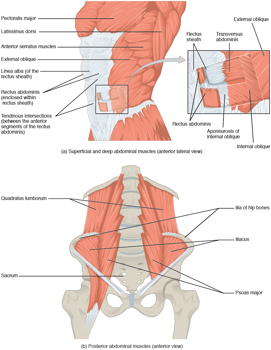
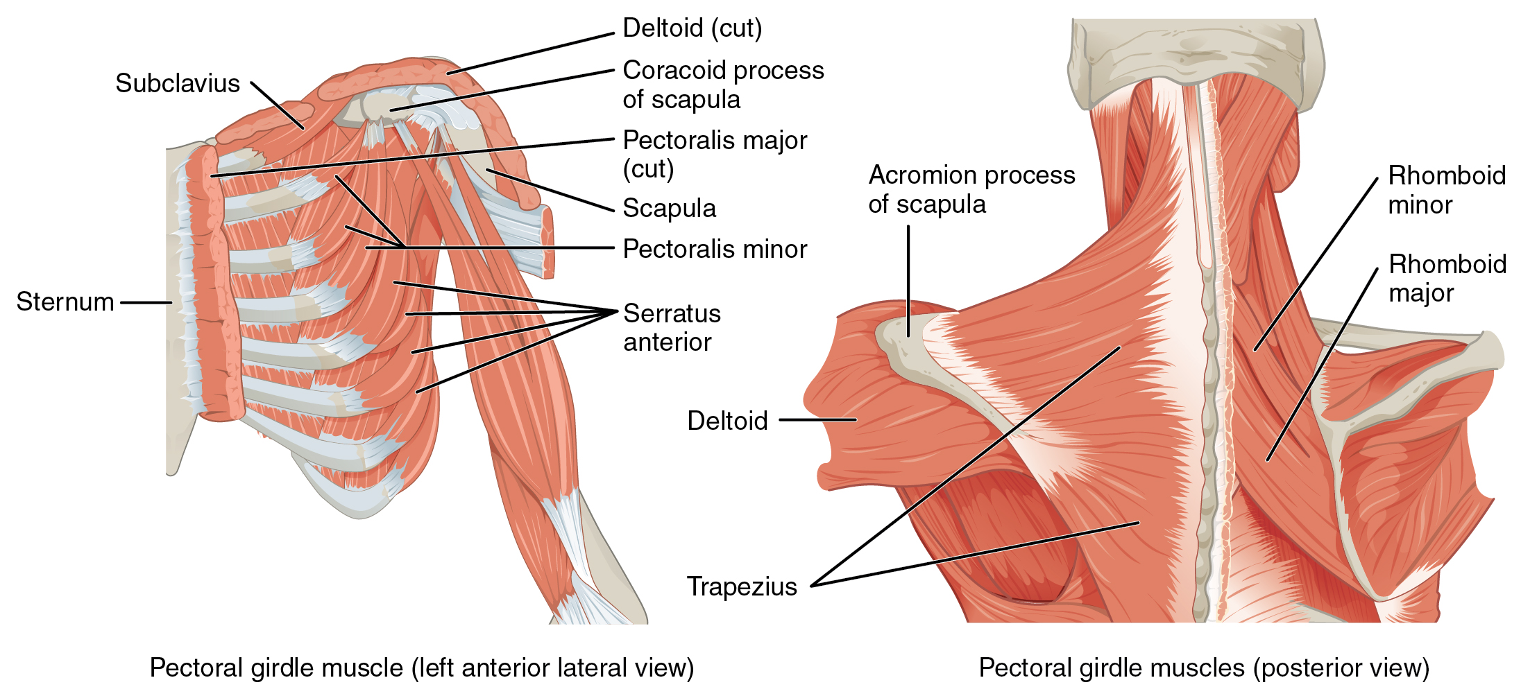
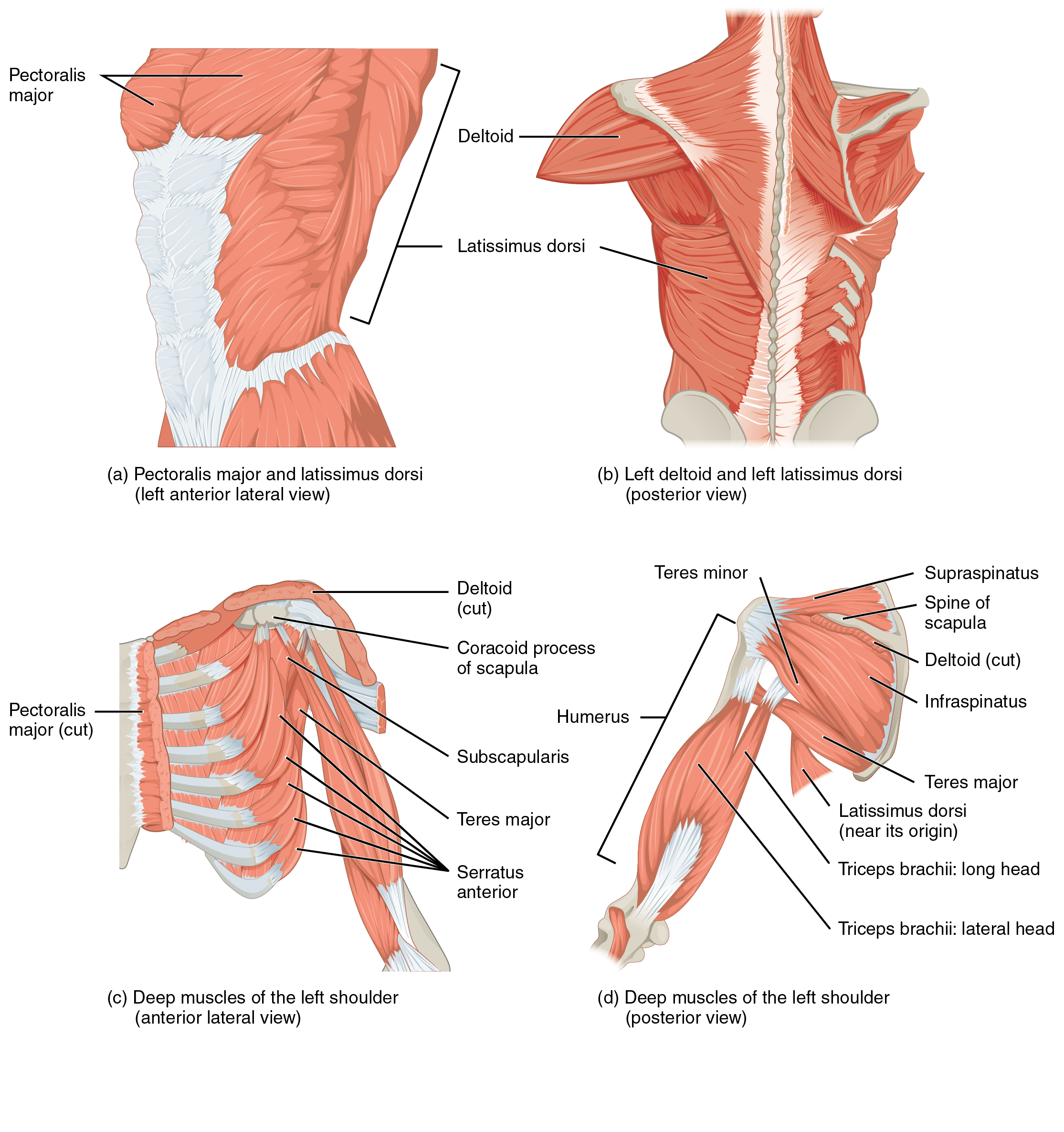
Pectoralis Major
| Muscle | Origin | Insertion | Innervation | Action |
|---|---|---|---|---|
| Pectoralis major | Clavicle (medial half) Sternum Costal cartilages 1-6 Rectus sheath (anterior layer) |
Humerus (crest of greater tubercle) | Lateral pectoral n. Medial pectoral n. C5 - T1 |
Entire muscle: Adduction, IR Clavicular & Sternocostal parts: Flexion, Aids in respiration when shoulder is fixed |
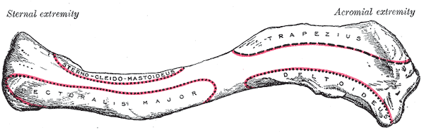
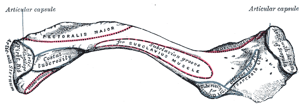
Origin
- Clavicular part: Clavicle (medial half)5
- Sternocostal part: sternum, costal cartilages 1–65
- Abdominal part: rectus sheath (anterior layer)5
Note
Manubrium and the aponeurosis of the external oblique are potential originations of the pectoralis major
Insertion
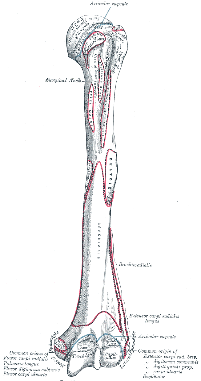
Humerus (crest of greater tubercle)5
Innervation
Action
- Entire muscle: GHJ Adduction, Internal rotation5
- Clavicular and sternocostal parts: Flexion; assist in respiration when shoulder is fixed5
Entire muscle: GHJ Adduction, Internal rotation
Clavicular and sternocostal parts: Flexion; assist in respiration when shoulder is fixed
Length test
- Pectoralis major contracture test (hands behind head)
Muscle-to-tendon Surgery
- Worse prognosis than tendon-to-bone because of slow healing
Tendon-to-bone Surgery
considerations
- Thoracic spine mobility
- Grip strength
- light cardio
- contralateral limb training (go light just to be safe to avoid irradiation)
Exercises
- Elbow CARs
- Spine CARs
Strain-Counterstrain
References
1.
Betts JG, Blaker W. Openstax Anatomy and Physiology. 2nd ed. OpenStax; 2022. https://openstax.org/details/books/anatomy-and-physiology-2e/?Book%20details
2.
Donnelly JM, Simons DG, eds. Travell, Simons & Simons’ Myofascial Pain and Dysfunction: The Trigger Point Manual. Third edition. Wolters Kluwer Health; 2019.
3.
Neumann DA, Kelly ER, Kiefer CL, Martens K, Grosz CM. Kinesiology of the Musculoskeletal System: Foundations for Rehabilitation. 3rd ed. Elsevier; 2017.
4.
Weinstock D. NeuroKinetic Therapy: An Innovative Approach to Manual Muscle Testing. North Atlantic Books; 2010.
5.
Gilroy AM, MacPherson BR, Wikenheiser JC, Voll MM, Wesker K, Schünke M, eds. Atlas of Anatomy. 4th ed. Thieme; 2020.
6.
Gray H. Anatomy of the Human Body. 20th ed. (Lewis WH, ed.). Lea & Febiger; 1918. https://www.bartleby.com/107/
7.
Myers HL, Devine WH, Fossum C, et al. Compendium Edition: Clinical Application of Counterstrain. Compendium ed. Osteopathic Press; 2012.
Citation
For attribution, please cite this work as:
Yomogida N, Kerstein C. Pectoralis Major. https://yomokerst.com/The
Archive/Anatomy/Skeletal Muscles/Upper Limb Muscles/Axioappendicular
Anterior Muscles/pectoralis_major.html