Scapula (Bone)
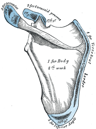
Osteologic features
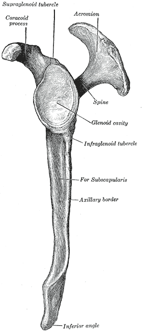
- Angles
- Borders
- Supraspinous fossa
- Infraspinous fossa
- Spine of scapula
- Root of the spine
- Acromion
- Clavicular facet
- Glenoid fossa
- Supraglenoid tubercle
- Infraglenoid tubercles
- Coracoid process
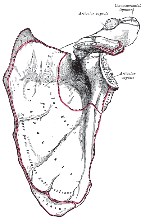
- Subscapular fossa
Superior angle
Lateral angle
Inferior angle
Superior border
One of the borders of the thoracic outlet2
Medial (vertebral) border
Lateral (axillary) border
Supraspinous fossa
Infraspinous fossa
Spine of Scapula
“The spine of the scapula provides a continuous line of attachments for the supporting trapezius muscle along its upper border, whereas the deltoid muscle, which suspends the humerus, gains origin from its lower border.”2
Root of the Spine of the Scapula
Acromion
- Part of the Acromioclavicular joint
“A prominent feature of the scapula is the large overhanging acromion (see Fig. 16-3), which, along with the coracoacromial ligament and the previously mentioned labrum, functionally enlarges the G-H socket. The position of the acromion also places the deltoid muscle in a dominant position to provide muscular support during elevation of the arm. The morphology of the acromion is variable with three types having been described—(1) a relatively flat undersurface (Type I), (2) a slightly convex undersurface (Type II), and (3) a hooked shape (Type III), with a 2018 study finding that individuals with a type III acromion had almost three times greater odds for an RTC tear as compared to those with a type I or II, concluding that a larger acromial index is associated with a greater likelihood of an RTC tear.9 This despite an earlier study in 2011 that concluded that the type of acromion was not found to have an important role in the etiology of chronic impingement syndrome.10”2
Clavicular facet
Glenoid fossa
Supraglenoid tubercle
Infraglenoid tubercle
Coracoid process
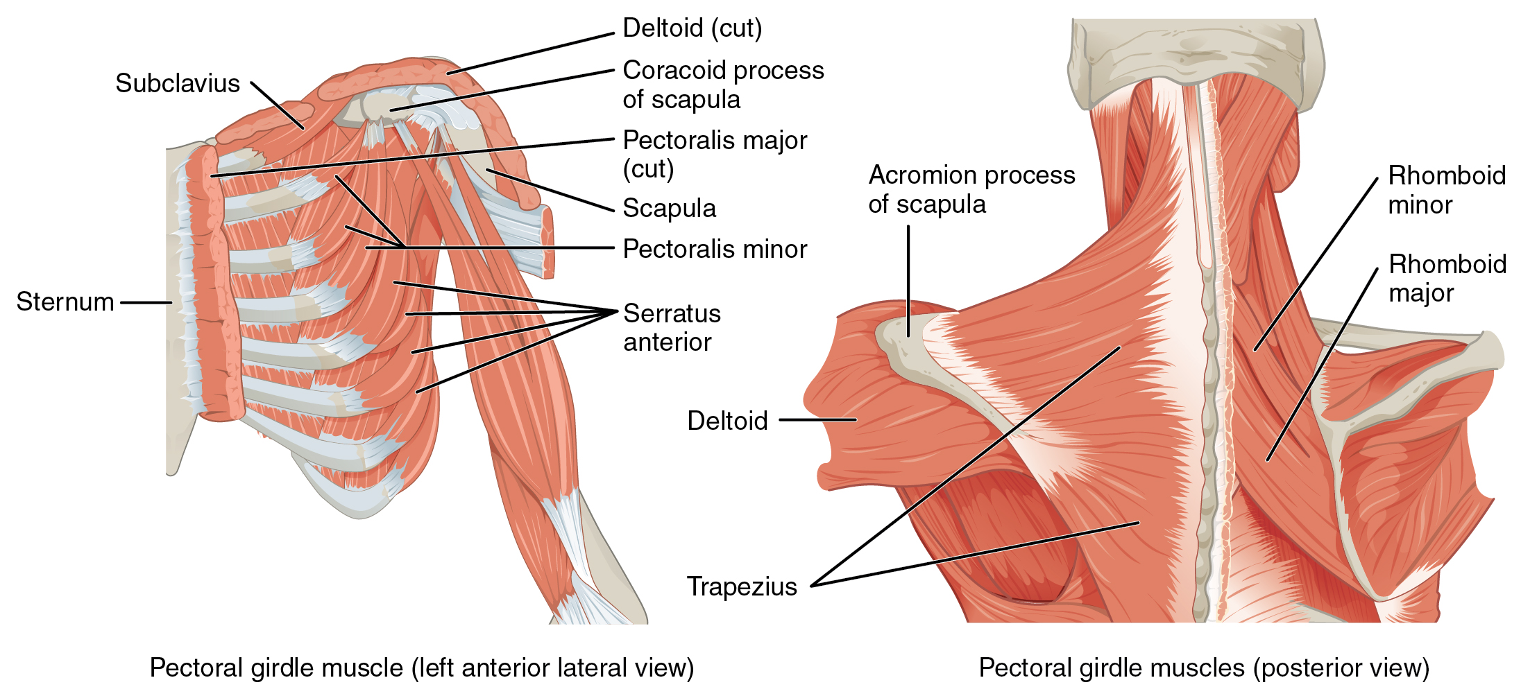
“The coracoid process (see Fig. 16-3) projects forward like a crow’s beak, for which it is named. This forward position provides an efficient lever whereby the small pectoralis muscle can help to stabilize the scapula. In addition, the process serves as a point of origin for the coracobrachialis and the short head of the biceps muscle”2
- Attachment for coracoclavicular trapezius ligament4
- Attachment for coracoclavicular conoid lig.4
The coracobrachialis and pectoralis minor both have insertions on the coracoid process, which creates a myofascial line5. When the arm is relaxed by one’s side since the pec minor and coracobrachialis fascial lines run in two different directions, thus the line is inactive5. However, when shoulder is brought into overhead flexion (i.e. tennis serve or hanging from a bar)5.