Serratus Anterior
| Muscle | Origin | Insertion | Innervation | Action |
|---|---|---|---|---|
| Serratus anterior | 1-9 Ribs | Superior angle of Scapula (costal and dorsal surfaces) Medial border of scapula (costal surface) Medial border of scapula (costal surface) Inferior angle of Scapula (costal & dorsal surfaces) |
Long thoracic n. C5 - C7 |
Superior part: Lowers raised arm Entire mm.: Protraction, Rib elevation (with fixed shoulder) Inferior part: Rotates scapula laterally forward |
Overview
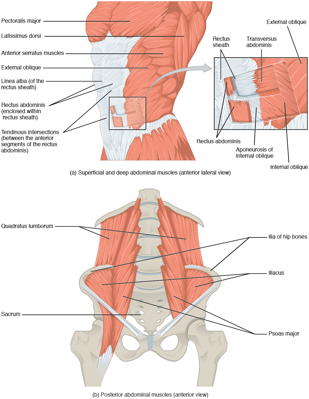
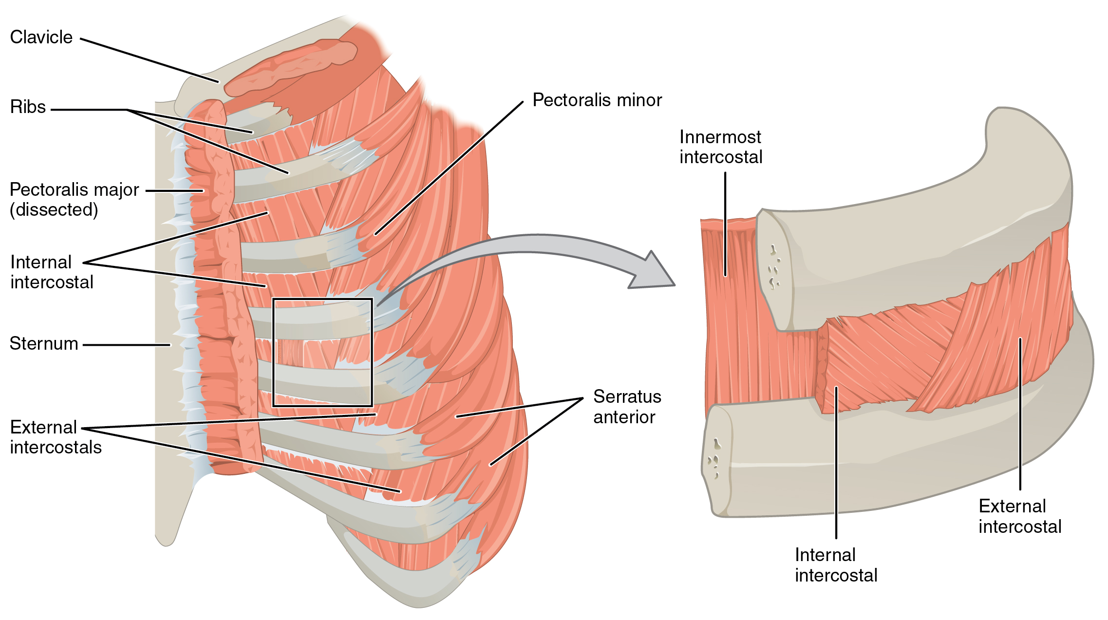
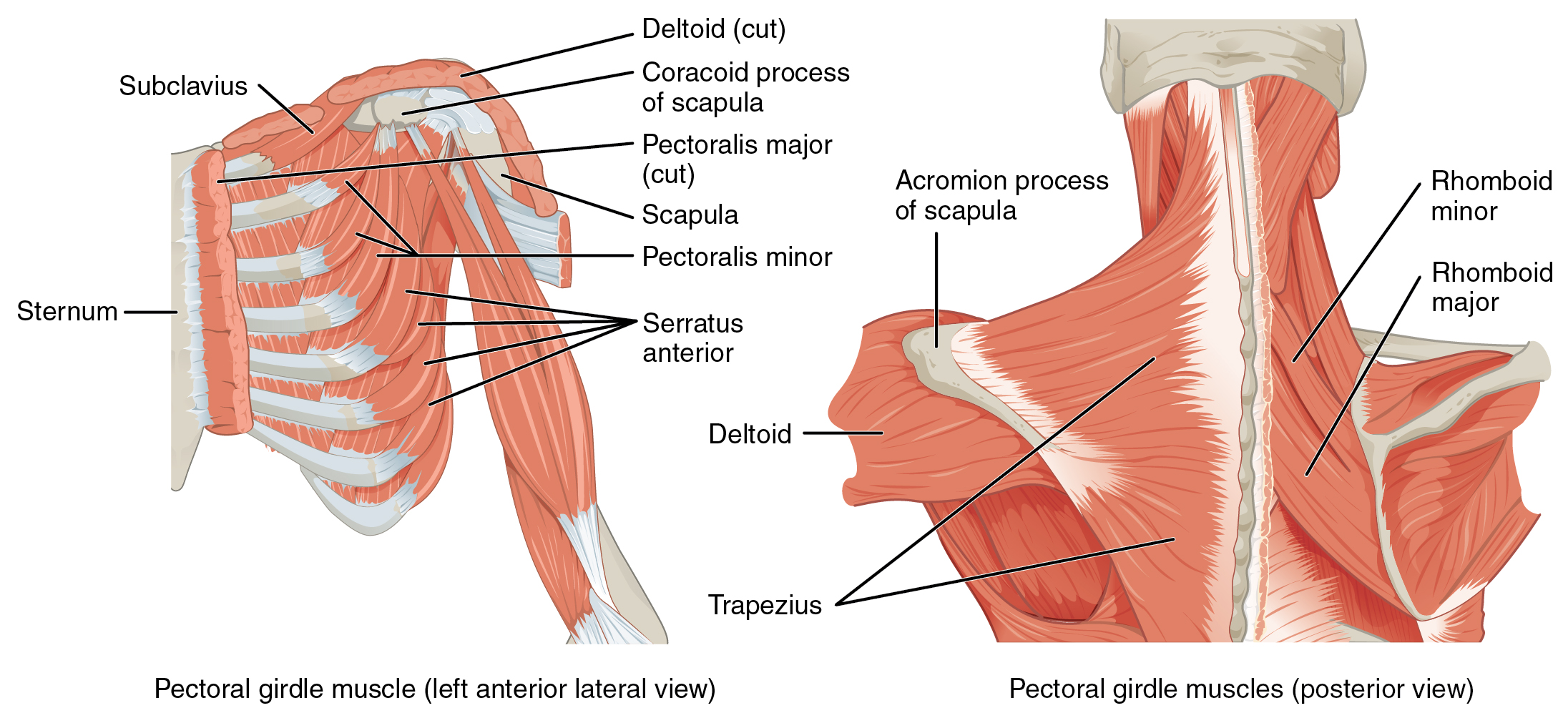
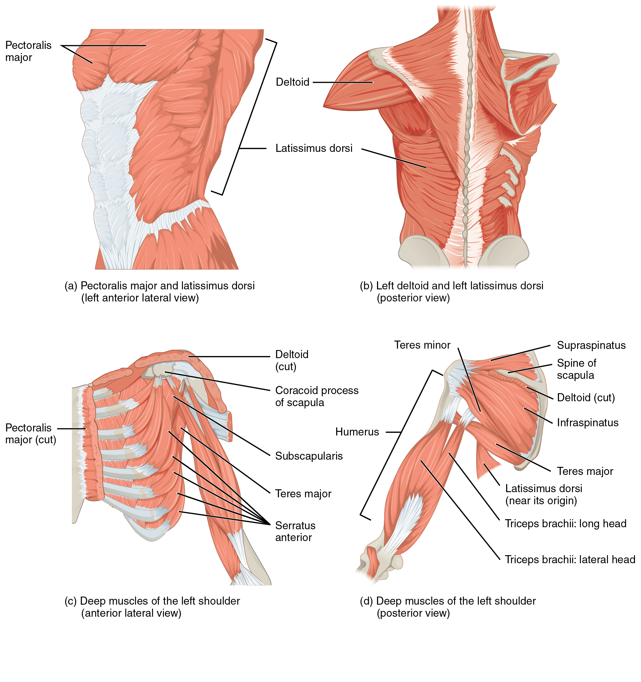
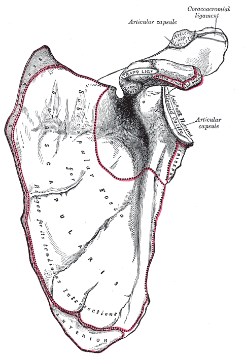
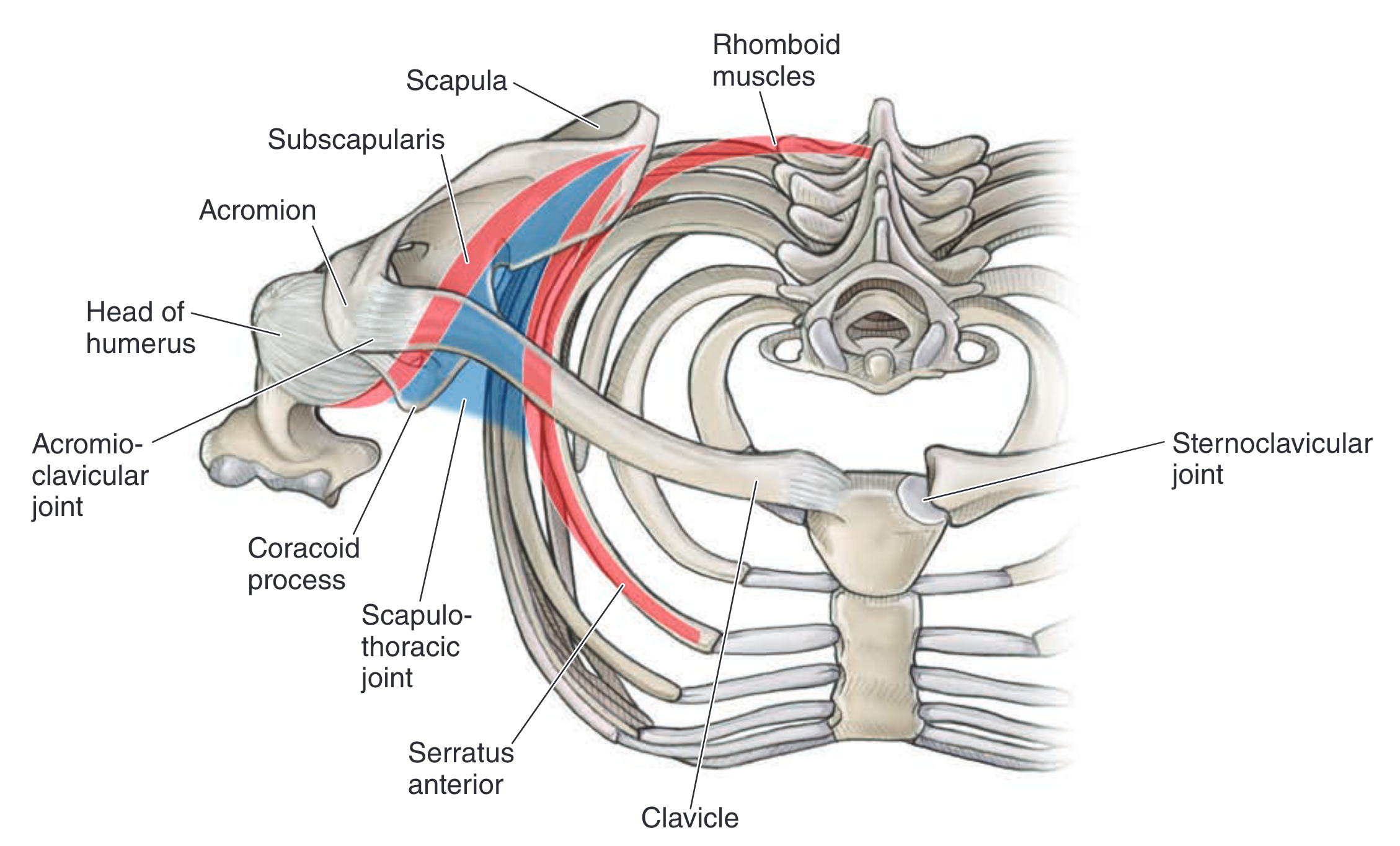
The serratus anterior is a fan-shaped muscle that spans the lateral wall of the thorax6. Most of the muscle belly lies deep to the scapula. The serratus inserts on the ribs, creating a sawtooth or “serrated” appearance6.
Origin
Insertion
- Superior part: Superior angle of Scapula (costal and dorsal surfaces)7
- Intermediate part: Medial border of Scapula (costal surface)7
- Inferior part: Medial border of Scapula (costal surface), Inferior angle (costal and dorsal surfaces)7
Nerve
The Serratus Anterior is solely innervated by the Long Thoracic N. (C5, C6, C7)7
Action
- Entire muscle:
- Superior part: Lowers the raised arm7 by depressing the scapula6.
- Inferior part: Rotates inferior angle of scapula laterally forward (allows elevation of arm above 90°)7
The superior and inferior parts of the serratus anterior have antagonistic actions in the vertical plane, which is why when the entire muscle activates, the scapula has no vertical translation.
The serratus anterior functions to stabilize the scapula, which provides proximal stability for the shoulder joint6.
Function
The Serratus Anterior (along with the rhomboids) serves to aid in scapular stability during arm elevation9
Release
Prone release
- Support the anterior shoulder with one arm
- Start at the lateral border of the scapula and palpate through to feel the serratus anterior’s origin on ribs 1-96.
- Press on the muscle on the rib.
- Do not press between the ribs
Paralysis
Causes:
- SCI
- peripheral nerve injury
- Nerve root?
- botox?
Serratus anterior paralysis can result in detachment of the scapula from the thorax known as “scapular winging.”