S2 Nerve Root (S2)
Peripheral Nerve Supply
| Nerve | Roots | Motor Innervation | Cutaneous Innervation |
|---|---|---|---|
| Posterior Cutaneous Nerve of the Thigh | |||
| Sciatic N. | |||
| Common Fibular N. | |||
| Tibial N. | |||
| Pudendal N. |
Muscle Supply
Motor Innervation
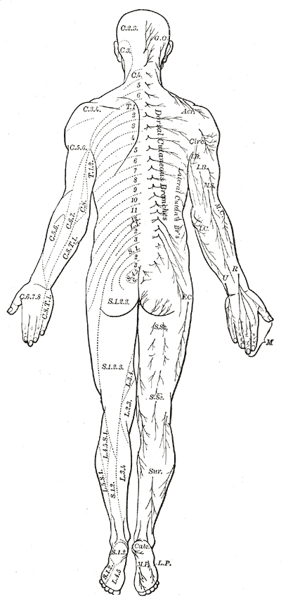
Dermatome

Examination
DTR
- Achilles Reflex (S1, S2)
References
1.
Gray H. Anatomy of the Human Body. 20th ed. (Lewis WH, ed.). Lea & Febiger; 1918. https://www.bartleby.com/107/
Citation
For attribution, please cite this work as:
Yomogida N, Kerstein C. S2 Nerve Root (S2). https://yomokerst.com/The
Archive/Anatomy/Nerve Roots/S2_nerve_root.html