Piriformis Muscle
| Muscle | Origin | Insertion | Innervation | Action |
|---|---|---|---|---|
| Piriformis | Anterior Sacrum | Apex of the Greater Trochanter | Sacral Plexus Direct Br. S1 - S2 |
Hip (at <60° flexion): ER Hip: Abduction, Extension, Stabilization Hip (at ≥60° flexion): IR |
Origin
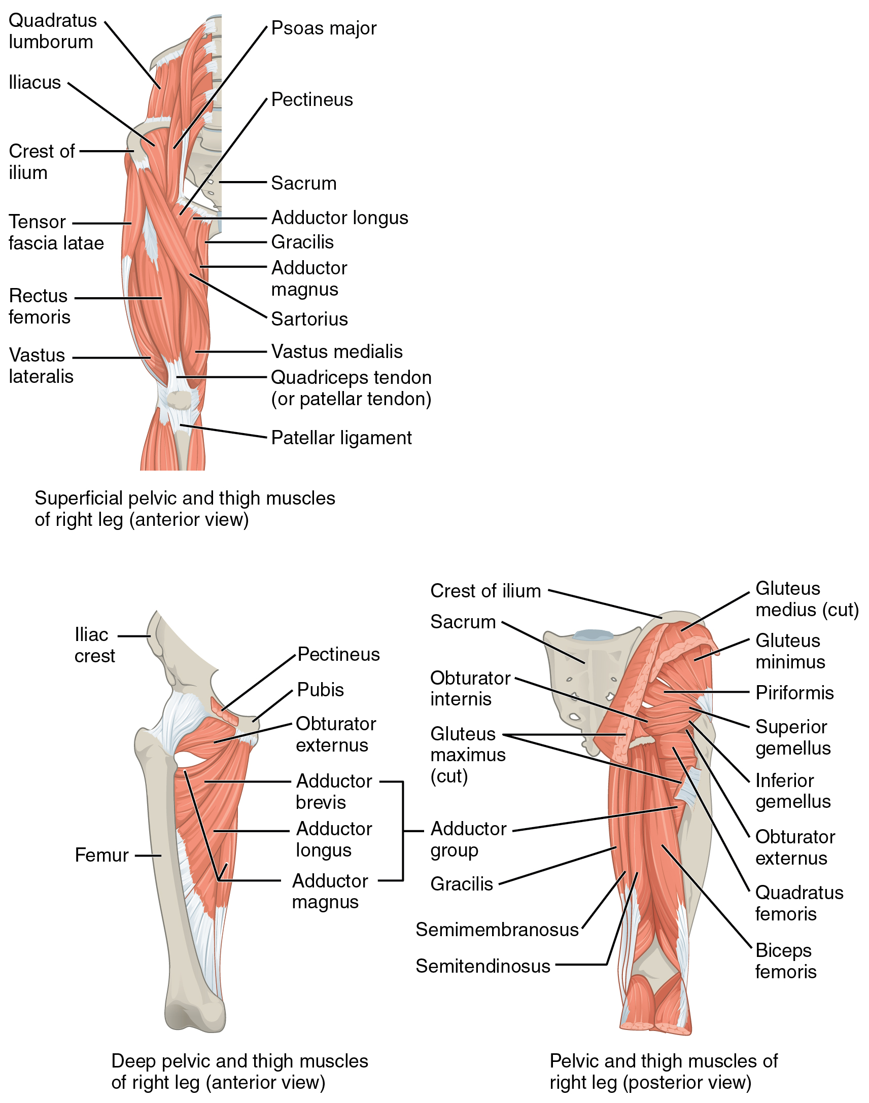
Anterior surface of sacrum6 from the 2nd-4th sacral segments7.
Path
After its origin the piriformis runs through the greater sciatic foramen before reaching its insertion on the greater trochanter7.
Insertion
Apex of the greater trochanter of the femur6.
Innervation
Action
According to Gilroy6, the piriformis functions in External rotation, abduction, extension and stabilization of the hip joint.
This is only partly true.
At ≥60° hip flexion, the piriformis produces an internal rotation torque4.
Neutral
- ER
- Flexion
- Abduction
60° Flexion
- IR
- Extension
- Abductor
Anatomy
The Sciatic n. runs superficial to all of the deep external rotators except for the piriformis8. The sciatic nerve can either runs deep to the piriformis and can exit inferiorly, through the piriformis muscle belly, or superior to the piriformis. As a result, overactivation of the piriformis can lead to neurogenic pain and symptoms and is termed Piriformis syndrome
Palpation
- Place the patient in prone8.
- Palpate the: coccyx, PSIS, and greater trochanter8.
- The coccyx to the PSIS is the superior and inferior bounds of the piriformis origin and the greater trochanter is the insertion8.
- Bisect the coccyx and PSIS and place a finger there8.
- Place your fingers along the imaginary line from the bisection to the greater trochanter8.
- Work through the superficial gluteus maximus to palpate the slender piriformis muscle belly8.
- Strum across the piriformis muscle belly to palpate its location8.
The sciatic nerve runs through this area, so be mindful of this when palpating.
Pathologies
- Piriformis Syndrome (Piriformis caused sciatica)
- Piriformis Tendinitis
Piriformis tendinitis
“Tenderness to deep palpation near the hook of the greater trochanter. Pain reproduced by piriformis stretch”9
Piriformis Syndrome
The Sciatic n. runs superficial to all of the deep external rotators except for the piriformis8. The sciatic nerve can either runs deep to the piriformis and can exit inferiorly, through the piriformis muscle belly, or superior to the piriformis. As a result, overactivation of the piriformis can lead to neurogenic pain and symptoms and is termed Piriformis syndrome
- Active piriformis test
- Seated piriformis test
Evaluation
- Active piriformis test
- Seated piriformis test
Active Release Technique (ART)
Active release technique for piriformis by Dr. Nick Perkins10
- Patient in contralateral sidelying
- STM to piriformis
- move from hip IR/ABD/Ext into hip ER/Adduction/Flexion
Manual Therapy
- Piriformis muscle technique11
Exercises
- FAddER Contract-Relax
- Therapist assisted
- Self-Stretch