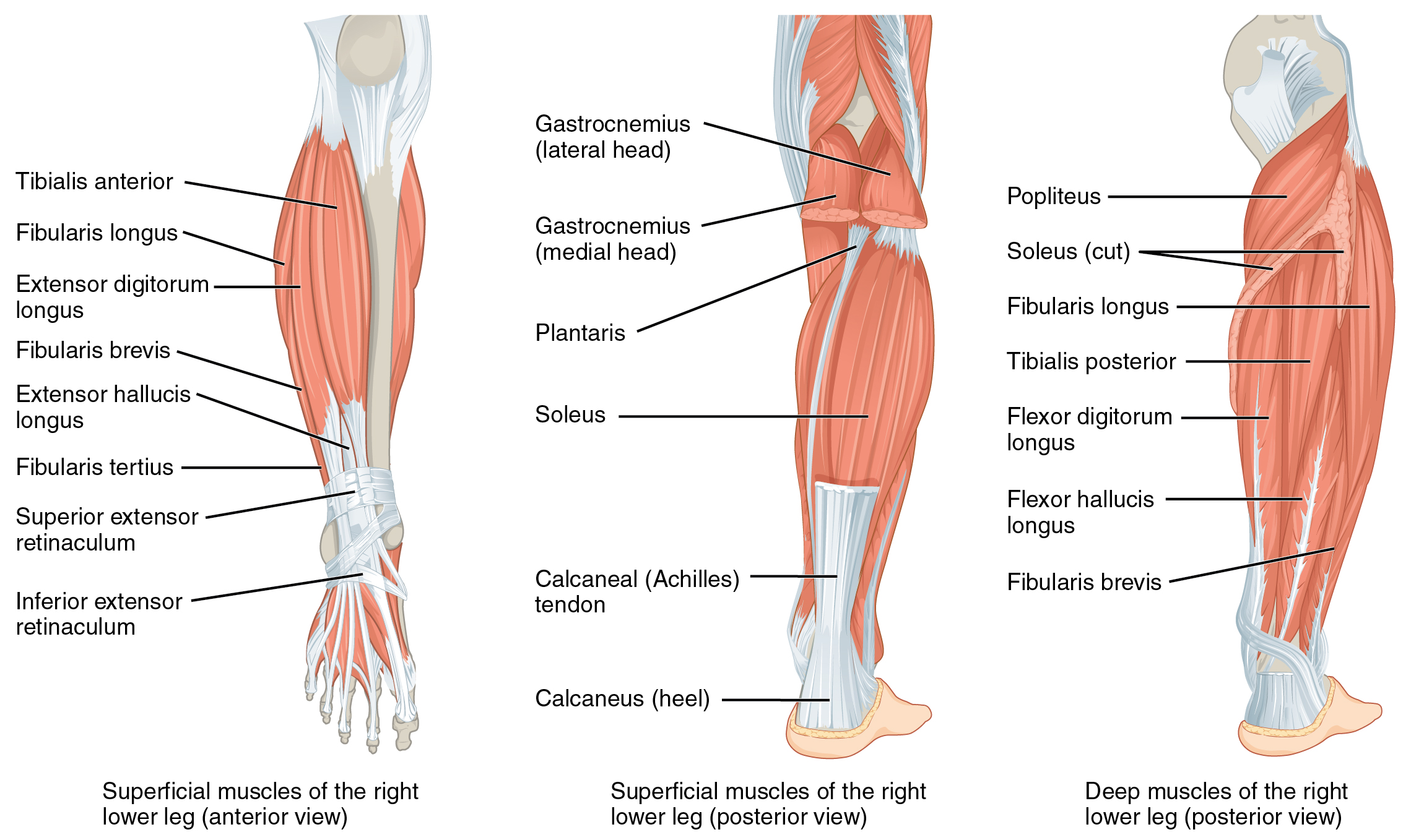
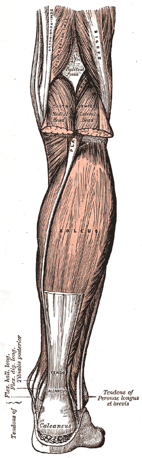
Soleus Muscle

| Muscle | Origin | Insertion | Innervation | Action |
|---|---|---|---|---|
| Soleus | Posterior Fibula Posterior Tibia |
Calcaneal tuberosity via Achilles' Tendon | Tibial n. S1 - S2 |
TCJ: PF Lower leg: Stabilization |
Origin
Insertion
Calcaneal tuberosity via the Achilles’ tendon6
Innervation
Action
Biomechanics
The Triceps Surae inserts onto the calcaneal tuberosity via the achilles tendon. The fulcrum is technically the distal foot and the external load acting upon this muscle, joint, tendon complex is at the tibia. Thus, this is a second class lever and as a result, the triceps surae create more torque relative to their force compared to most other muscles in the body.
Muscle Length Test
- Test DF with Knee flexed to 45°
Strength Testing
“The soleus muscle produces plantar flexion of the ankle joint, regardless of the position of the knee. To determine the individual functioning of the soleus as a plantar flexor, the knee is flexed to minimize the effect of the gastrocnemius muscle. The soleus is tested in a similar manner to that of the gastrocnemius, except that the patient performs the unilateral heel raise with some degree of knee flexion. Ability to perform 10–15 raises in this fashion is considered normal, 5-9 raises graded as fair, 1–4 raises graded as poor, and zero repetitions graded as nonfunctional. Alternatively, the strength of the soleus can be tested with the patient in prone”
Pails & Rails
P.A.I.L.’s
- Plantarflexion
R.A.I.L.’s
- Dorsiflexion
Release
Releasing the soleus at its origin can be important for freeing the tibial nerve as it passes through the soleus.
- Patient in prone
- Palpate lateral gastrocnemius head
- Move medially to palpate the tibial nerve
- Follow the tibial nerve inferiorly until you reach the soleus’ origin.
- Apply pure pressure to release the soleus
Use the SLR Ankle contract-relax as a test-retest and a way to glide the tibial nerve.