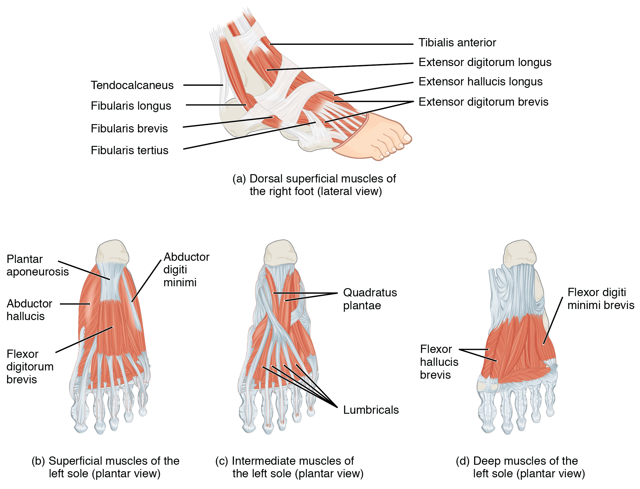
Flexor Digitorum Brevis (FDB)
Overview
| Muscle | Origin | Insertion | Innervation | Action |
|---|---|---|---|---|
| Flexor digitorum brevis | Medial tubercle of calcaneal tuberosity Plantar aponeurosis |
Sides of middle phalanx 2-5 | Medial Plantar n. S1 - S2 |
2nd-5th Toe: MTP Flexion, PIP Flexion Arch support: Longitudinal arch support |
Origin
Insertion
Sides of middle phalanges #2-55
Innervation
Action
MMT
“The FDL and brevis muscles produce IP joint flexion. The motion is tested with the foot in the anatomic position. If the gastrocnemius muscle is shortened, preventing the ankle from assuming the anatomic position, the knee is flexed. The toes may be tested simultaneously. The foot is held in the midposition, and the metatarsals are stabilized. Resistance is applied beneath the distal and proximal phalanges”6
References
1.
Betts JG, Blaker W. Openstax Anatomy and Physiology. 2nd ed. OpenStax; 2022. https://openstax.org/details/books/anatomy-and-physiology-2e/?Book%20details
2.
Gray H. Anatomy of the Human Body. 20th ed. (Lewis WH, ed.). Lea & Febiger; 1918. https://www.bartleby.com/107/
3.
Donnelly JM, Simons DG, eds. Travell, Simons & Simons’ Myofascial Pain and Dysfunction: The Trigger Point Manual. Third edition. Wolters Kluwer Health; 2019.
4.
Neumann DA, Kelly ER, Kiefer CL, Martens K, Grosz CM. Kinesiology of the Musculoskeletal System: Foundations for Rehabilitation. 3rd ed. Elsevier; 2017.
5.
Gilroy AM, MacPherson BR, Wikenheiser JC, Voll MM, Wesker K, Schünke M, eds. Atlas of Anatomy. 4th ed. Thieme; 2020.
6.
Dutton M. Dutton’s Orthopaedic Examination, Evaluation, and Intervention. 5th ed. McGraw Hill Education; 2020.
Citation
For attribution, please cite this work as:
Yomogida N, Kerstein C. Flexor Digitorum Brevis
(FDB). https://yomokerst.com/The
Archive/Anatomy/Skeletal Muscles/Lower limb muscles/Ankle and
Foot/Intrinsic Dorsal Foot
Muscles/flexor_digitorum_brevis.html