Flexor Hallucis Longus (FHL)
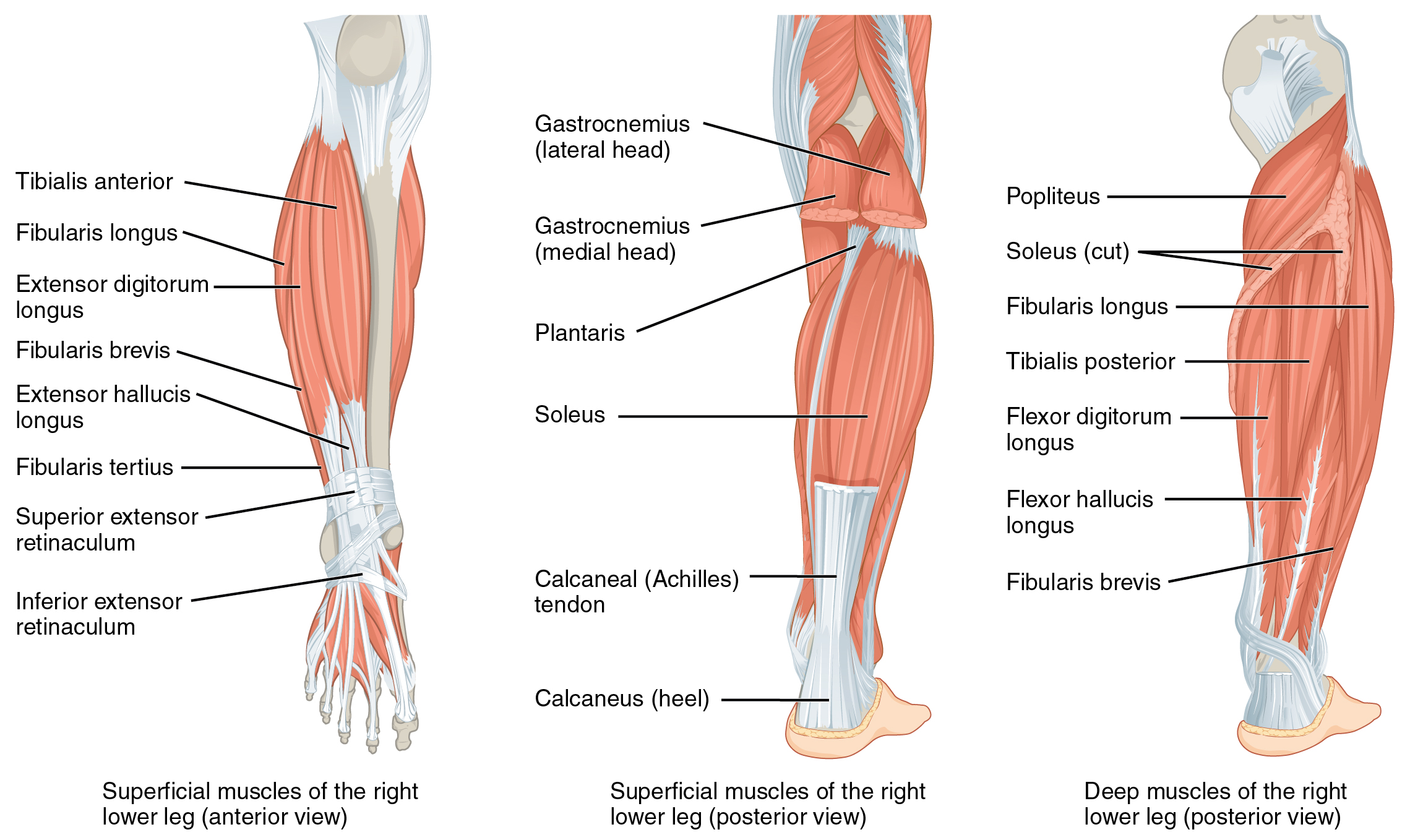
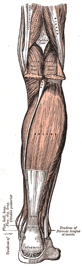

Overview
| Muscle | Origin | Insertion | Innervation | Action |
|---|---|---|---|---|
| Flexor hallucis longus | Posterior distal 2/3 of Fibula IO membrane |
Base of 1st Distal Phalanx | Tibial n. L5 - S2 |
TCJ: PF STJ: Inversion 1st Toe: MTP Flexion, IP Flexion |
Origin
Insertion
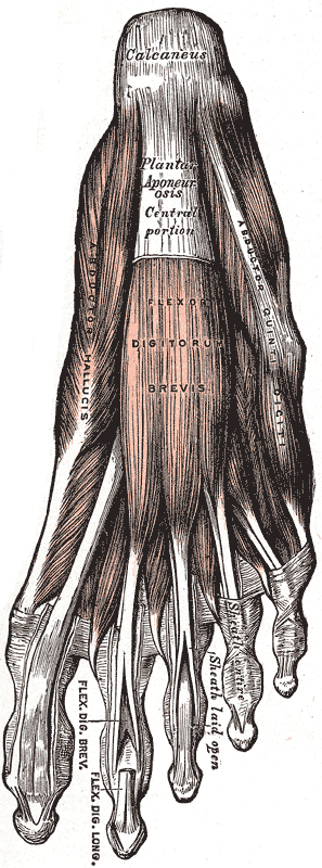
1st distal phalanx (base)6
Innervation
Action
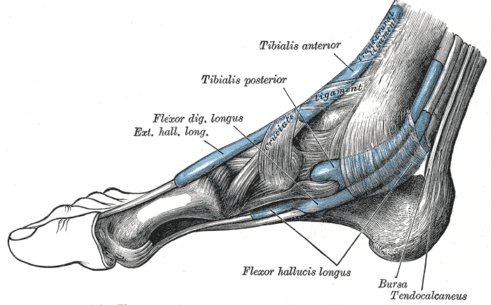
The posterior tibialis and flexor hallucis longus also improves the stability of ankle joint by creating a dynamic pinch between the malleoli, which improves joint contact during plantarflexio7.
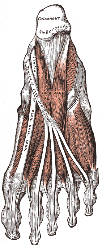
The FHL also plays a role in calcaneal frontal plane stabilization. Although it does not insert on the calcaneus, the FHL tendon “hooks” onto the calcaneus in the sustentaculum tali allowing it to exert a force upon the calcaneus7. The FHL tendon can control and prevent excessive calcaneal varus/adduction.
MMT
“The FHB VIDEO and FHL muscles VIDEO produce MTP joint flexion and IP joint flexion. The foot is maintained in midposition. The first metatarsal is stabilized, and resistance is applied beneath the proximal and distal phalanx of the great toe into toe extension.”8
Pails & Rails
P.A.I.L.’s
- Plantarflexion
- Inversion
- 1st MTP/IP flexion
R.A.I.L.’s
- Dorsiflexion
- Eversion
- 1st toe MTP/IP extension