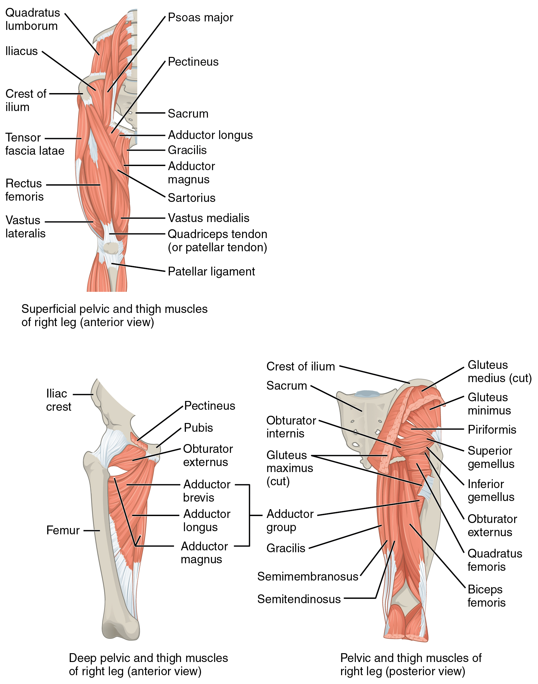
Semimembranosus

Authors
Affiliations

Doctor of Physical Therapy

B.S. in Kinesiology

Doctor of Physical Therapy

B.A. in Neuroscience

Figure 1: Hip and Thigh Muscles

Cross-section of middle thigh2

Cross-section of middle thigh
Figure 2: Muscles of the gluteal and posterior femoral region
Figure 3: Superficial posterior leg muscles
Reading list
  • Ch60 Hamstring muscles
  • Neuman
  • MMT
Muscle Origin Insertion Innervation Action

Origin

Ischial tuberosity

Insertion

  • Medial tibial condyle
  • Oblique popliteal lig.
  • Popliteus fascia

Innervation

Tibial N. (Tibial division of Sciatic N.) (L5, S1, S2).

Action

  • Hip: Extension
  • Pelvic Stabilization: Sagittal plane
  • Knee: Flexion and Internal rotation

Overview

“The semimembranosus attaches distally to the posterior side of the medial condyle of the tibia. Additional distal attachments of this muscle include the MCL, both menisci, and the oblique popliteal ligament. For most of its course, the sinewy semitendinosus tendon lies posterior to the semimembranosus muscle. Just proximal to the knee, however, the tendon of the semitendinosus courses anteriorly toward its distal attachment on the anteriormedial aspect of tibia. Both heads of the biceps femoris attach primarily to the head of the fibula, with lesser insertions to the lateral collateral ligament, the capsule of the proximal tibiofibular joint, and the lateral tubercle of the tibia.265”

“The semimembranosus muscle (Fig. 19-8) arises from the lateral facet of the ischial tuberosity and the ischial ramus. This muscle inserts into the posteromedial aspect of the medial tibial condyle and has a key expansion that reinforces the posteromedial corner of the knee capsule. The semimembranosus pulls the meniscus posteriorly, and internally rotates the tibia on the femur, during knee flexion, although its primary function is to extend the hip and flex the knee.”

References

1.
Betts JG, Blaker W. Anatomy and Physiology. 2nd ed. OpenStax; 2022. https://openstax.org/details/books/anatomy-and-physiology-2e/?Book%20details
2.
Gray H. Anatomy of the Human Body. 20th ed. (Lewis WH, ed.). Lea & Febiger; 1918. https://www.bartleby.com/107/
3.
Donnelly JM, Simons DG, eds. Travell, Simons & Simons’ Myofascial Pain and Dysfunction: The Trigger Point Manual. Third edition. Wolters Kluwer Health; 2019.
4.
Neumann DA, Kelly ER, Kiefer CL, Martens K, Grosz CM. Kinesiology of the Musculoskeletal System: Foundations for Rehabilitation. 3rd ed. Elsevier; 2017.
5.
Weinstock D. NeuroKinetic Therapy: An Innovative Approach to Manual Muscle Testing. North Atlantic Books; 2010.
6.
Gilroy AM, MacPherson BR, Wikenheiser JC, Voll MM, Wesker K, Schünke M, eds. Atlas of Anatomy. 4th ed. Thieme; 2020.
7.
Dutton M. Dutton’s Orthopaedic Examination, Evaluation, and Intervention. 5th ed. McGraw Hill Education; 2020.

Citation

For attribution, please cite this work as: