Semitendinosus
| Muscle | Origin | Insertion | Innervation | Action |
|---|---|---|---|---|
| Semitendinosus | Ischial tuberosity Sacrotuberous lig. (common head with biceps femoris long head) |
Pes anserine | Tibial n. L5 - S2 |
Hip: Extension Knee: Flexion, IR Pelvis: Sagittal stabilization |
Origin
- Ischial tuberosity6,7
- Sacrotuberous lig.6
- (common head with long head of biceps femoris)6
Insertion
- Medial to the tibial tuberosity in the Pes Anserine6
- (along with the tendons of gracilis and sartorius)6
Nerve
Action
Overview
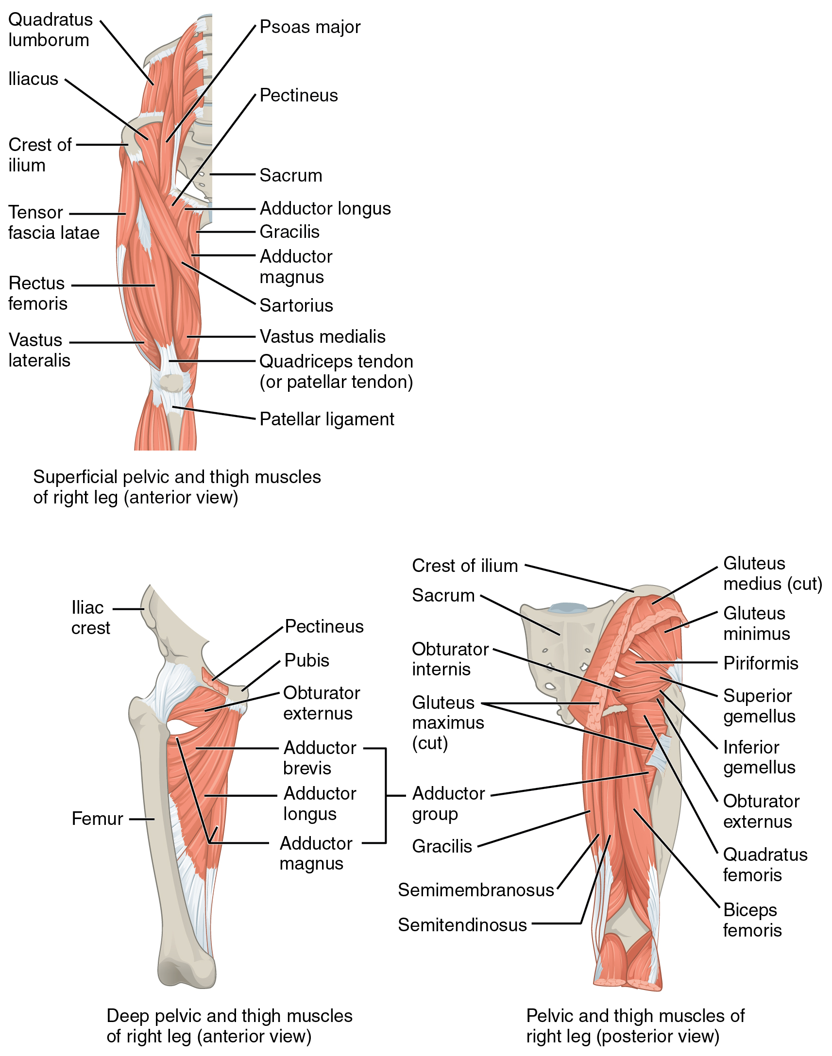
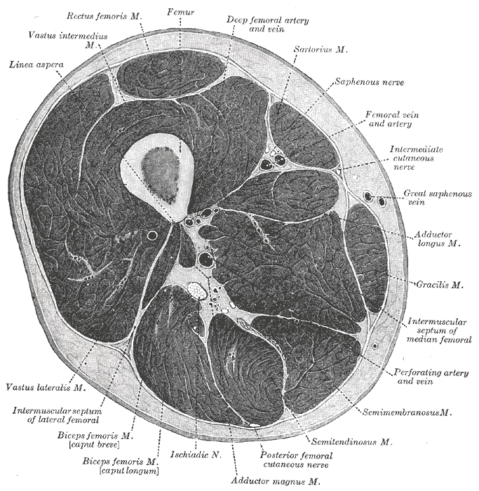
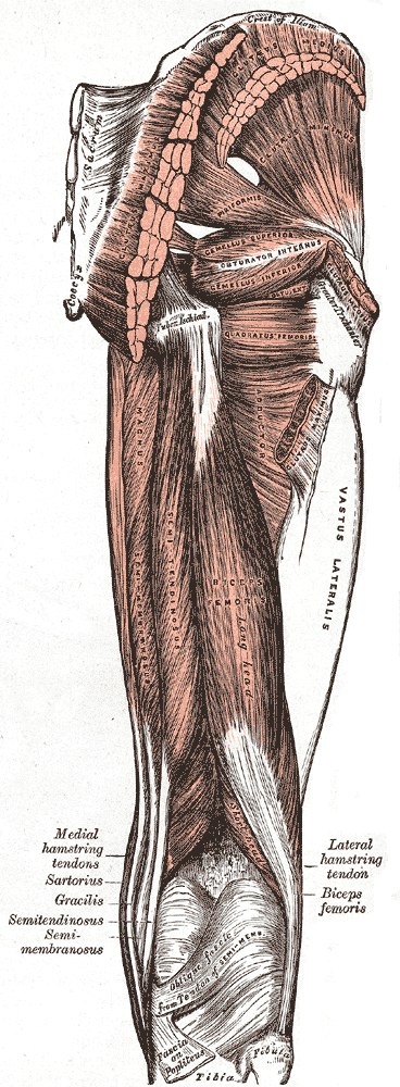
“The semitendinosus muscle (see Fig. 19-8) arises from the upper portion of the ischial tuberosity via a shared tendon with the long head of the biceps femoris. From there, it travels distally, becoming cord-like about two-thirds of the way down the posteromedial thigh. Passing over the MCL, it inserts into the medial surface of the tibia and deep fascia of the lower leg, distal to the gracilis attachment, and posterior to the sartorius attachment. These three structures are collectively called the pes anserinus (“goose foot”) at this point. Like the semimembranosus, the semitendinosus functions to extend the hip, flex the knee, and internally rotate the tibia.”8
References
1.
Betts JG, Blaker W. Openstax Anatomy and Physiology. 2nd ed. OpenStax; 2022. https://openstax.org/details/books/anatomy-and-physiology-2e/?Book%20details
2.
Gray H. Anatomy of the Human Body. 20th ed. (Lewis WH, ed.). Lea & Febiger; 1918. https://www.bartleby.com/107/
3.
Donnelly JM, Simons DG, eds. Travell, Simons & Simons’ Myofascial Pain and Dysfunction: The Trigger Point Manual. Third edition. Wolters Kluwer Health; 2019.
4.
Neumann DA, Kelly ER, Kiefer CL, Martens K, Grosz CM. Kinesiology of the Musculoskeletal System: Foundations for Rehabilitation. 3rd ed. Elsevier; 2017.
5.
Weinstock D. NeuroKinetic Therapy: An Innovative Approach to Manual Muscle Testing. North Atlantic Books; 2010.
6.
Gilroy AM, MacPherson BR, Wikenheiser JC, Voll MM, Wesker K, Schünke M, eds. Atlas of Anatomy. 4th ed. Thieme; 2020.
7.
Jones B. B Project Foundations. b Project; 2025.
8.
Dutton M. Dutton’s Orthopaedic Examination, Evaluation, and Intervention. 5th ed. McGraw Hill Education; 2020.
Citation
For attribution, please cite this work as:
Yomogida N, Kerstein C. Semitendinosus. https://yomokerst.com/The
Archive/Anatomy/Skeletal Muscles/Lower limb muscles/Thigh
Muscles/Posterior Thigh Muscles/semitendinosus.html


