Rectus Femoris Muscle
| Muscle | Origin | Insertion | Innervation | Action |
|---|---|---|---|---|
| Rectus femoris | AIIS Acetabular roof |
Tibial tuberosity via Patellar ligament | Femoral n. L2 - L4 |
Hip: Flexion Knee: Extension |
Origin
- Anterior inferior iliac spine (AIIS)4
- Acetabular roof of hip joint4
- Ilium5
The rectus femoris origin on the ASIS combines with the path of the ilio-femoral lig. strengthening the anterior aspect of the joint capsule5.
Insertion
Tibial tuberosity (via patellar lig.)4
Nerve
Femoral nerve (L2, L3, L4)4
Action
The rectus femoris can only generate 20% of the total force of the quadriceps group, but it cannot produce full knee extension in isolation5.
The rectus femoris muscle is a 2-joint muscle, acting on both the hip and the knee. As a result, these two joints can impact the rectus femoris’ efficiency on the other. The hip joint can significantly impact the rectus femoris’ role in the knee extensor mechanism6. In addition, the rectus femoris’ ability to efficiently flex the knee is proportional to the degree of hip flexion which we discuss more here5.
“The line of pull of the rectus femoris, with respect to the patella, is at an angle of about 5 degrees with the femoral shaft (see Fig. 20-12).”6
Biomechanics
- When in hip flexion, the ASIS and superior margin of the trochlea is shortened5.
- At hip neutral, the distance between the ASIS and superior margin of the trochlea increases5.
- In hip extension, the distance between the ASIs and superior margin of the trochlea is maximized5.
As the lower extremity moves into hip flexion and knee extension, the rectus femoris continues to shorten, resulting in active insufficiency, demonstrated as a decrease in knee extension efficiency. This decrease in efficiency is countered through an increase in vastus lateralis and vastus medialis recruitment5.
Force Couple
Erector Spinae and the Rectus Femoris are considered a force couple since both produce anterior pelvic tilt
Notes
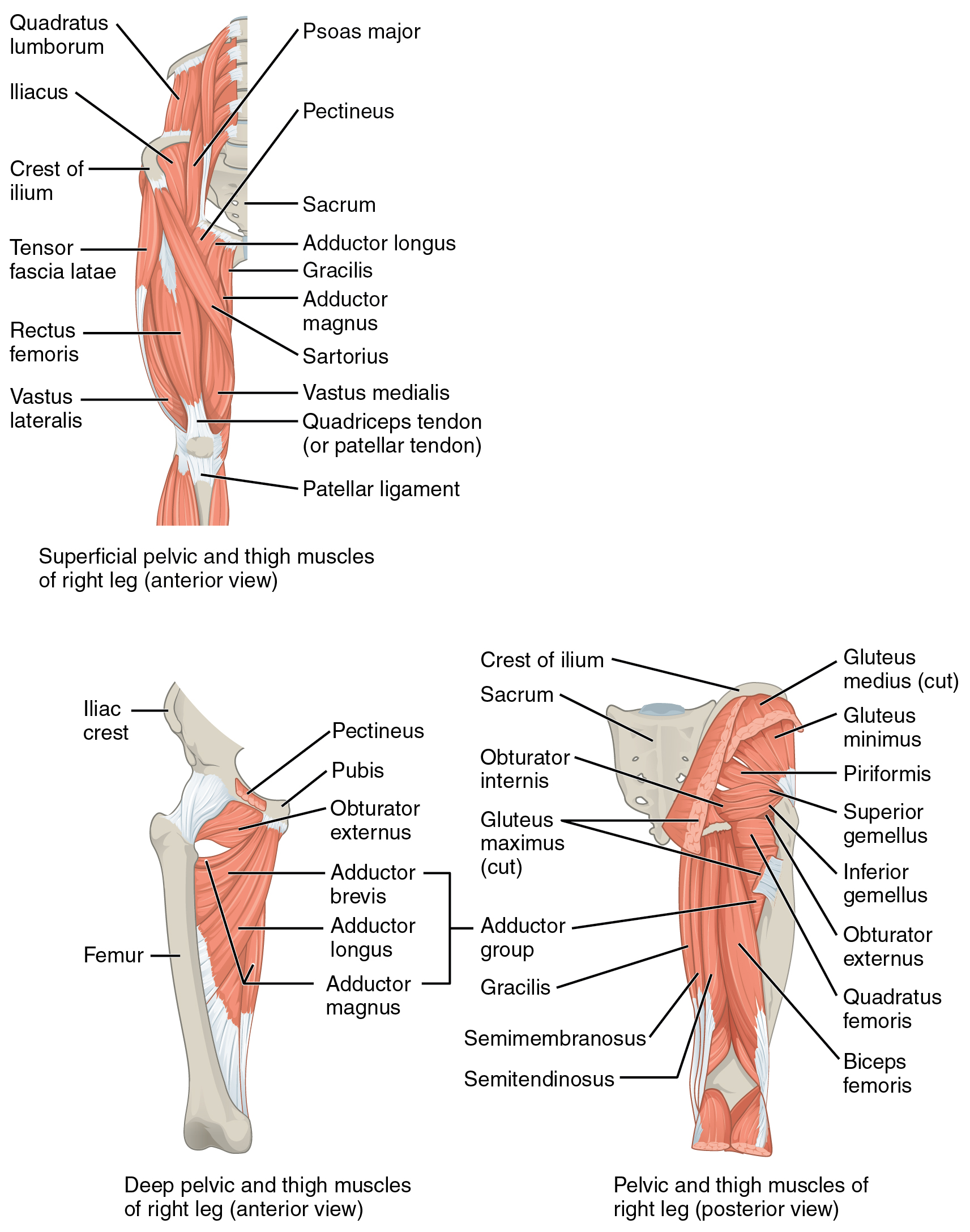
“The rectus femoris attaches to the pelvis near the anterior-inferior iliac spine and immediately superior to the acetabulum. The vastus muscles, however, attach to an extensive part of the femur, particularly the anterior-lateral shaft and the linea aspera (see Fig. 12.5). Although the vastus lateralis has the largest cross-sectional area of the quadriceps muscles, the vastus medialis extends farther distally toward the knee.”7
“The proximal part of the rectus femoris emerges between the limbs of an inverted V formed by the sartorius and tensor fasciae latae (see Fig. 12.26). This large bipennateshaped muscle has its proximal attachments on the anteriorinferior iliac spine, along the superior rim of the acetabulum, and in the adjacent joint capsule. The relatively robust capsular attachment made by the “reflected” tendon of the rectus femoris has been described as an important stabilizer of the anterior capsule.238 Along with the other members of the quadriceps, the rectus femoris attaches to the tibia via the patellar tendon. The rectus femoris is responsible for about onethird of the total isometric flexion torque at the hip.144 In addition, the rectus femoris is a primary knee extensor. The combined twojoint actions of this important muscle are considered in Chapter 13. The anatomy and function of the pectineus and adductor longus are described in the section on the adductors of the hip.”7