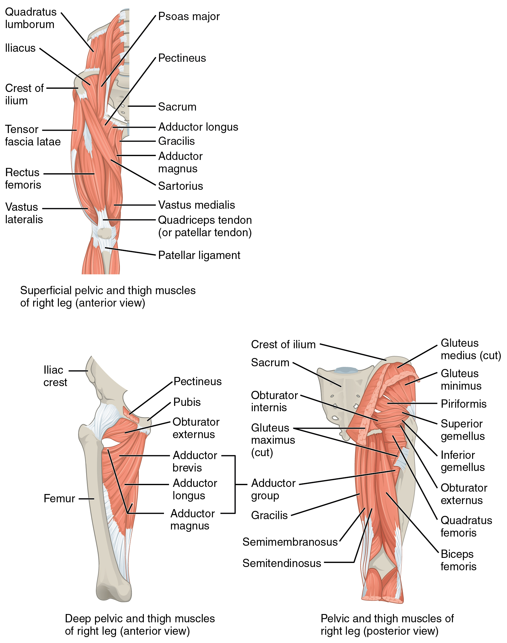
Vastus Lateralis (VL)
| Muscle | Origin | Insertion | Innervation | Action |
|---|---|---|---|---|
| Vastus lateralis | Lateral lip of Linea aspera Lateral surface of Greater trochanter |
Tibial tuberosity via Patellar ligament Tibial tuberosity and patella via Patellar retinacula |
Femoral n. L2 - L4 |
Knee: Extension |
Origin
Some sources suggest that the vastus lateralis originates from the lateral lip of the gluteal tuberosity as well5.
Insertion
Nerve
Femoral N. (L2, L3, L4)4
Action
Knee: extension4
It should be noted that the vastus medialis is stronger and descends further than the vastus lateralis5.
“The VL (Fig. 19-7) is composed of two functional parts: the VL and the vastus lateralis oblique (VLO). The VL has a line of pull of about 12–15° to the long axis of the femur in the frontal plane, whereas the VLO has a pull of 38–48°.”6
References
1.
Betts JG, Blaker W. Openstax Anatomy and Physiology. 2nd ed. OpenStax; 2022. https://openstax.org/details/books/anatomy-and-physiology-2e/?Book%20details
2.
Gray H. Anatomy of the Human Body. 20th ed. (Lewis WH, ed.). Lea & Febiger; 1918. https://www.bartleby.com/107/
3.
Weinstock D. NeuroKinetic Therapy: An Innovative Approach to Manual Muscle Testing. North Atlantic Books; 2010.
4.
Gilroy AM, MacPherson BR, Wikenheiser JC, Voll MM, Wesker K, Schünke M, eds. Atlas of Anatomy. 4th ed. Thieme; 2020.
5.
Jones B. B Project Foundations. b Project; 2025.
6.
Dutton M. Dutton’s Orthopaedic Examination, Evaluation, and Intervention. 5th ed. McGraw Hill Education; 2020.
Citation
For attribution, please cite this work as:
Yomogida N, Kerstein C. Vastus Lateralis (VL).
https://yomokerst.com/The
Archive/Anatomy/Skeletal Muscles/Lower limb muscles/Thigh
Muscles/Anterior Thigh Muscles/vastus_lateralis.html