Vastus Medialis (VM)
| Muscle | Origin | Insertion | Innervation | Action |
|---|---|---|---|---|
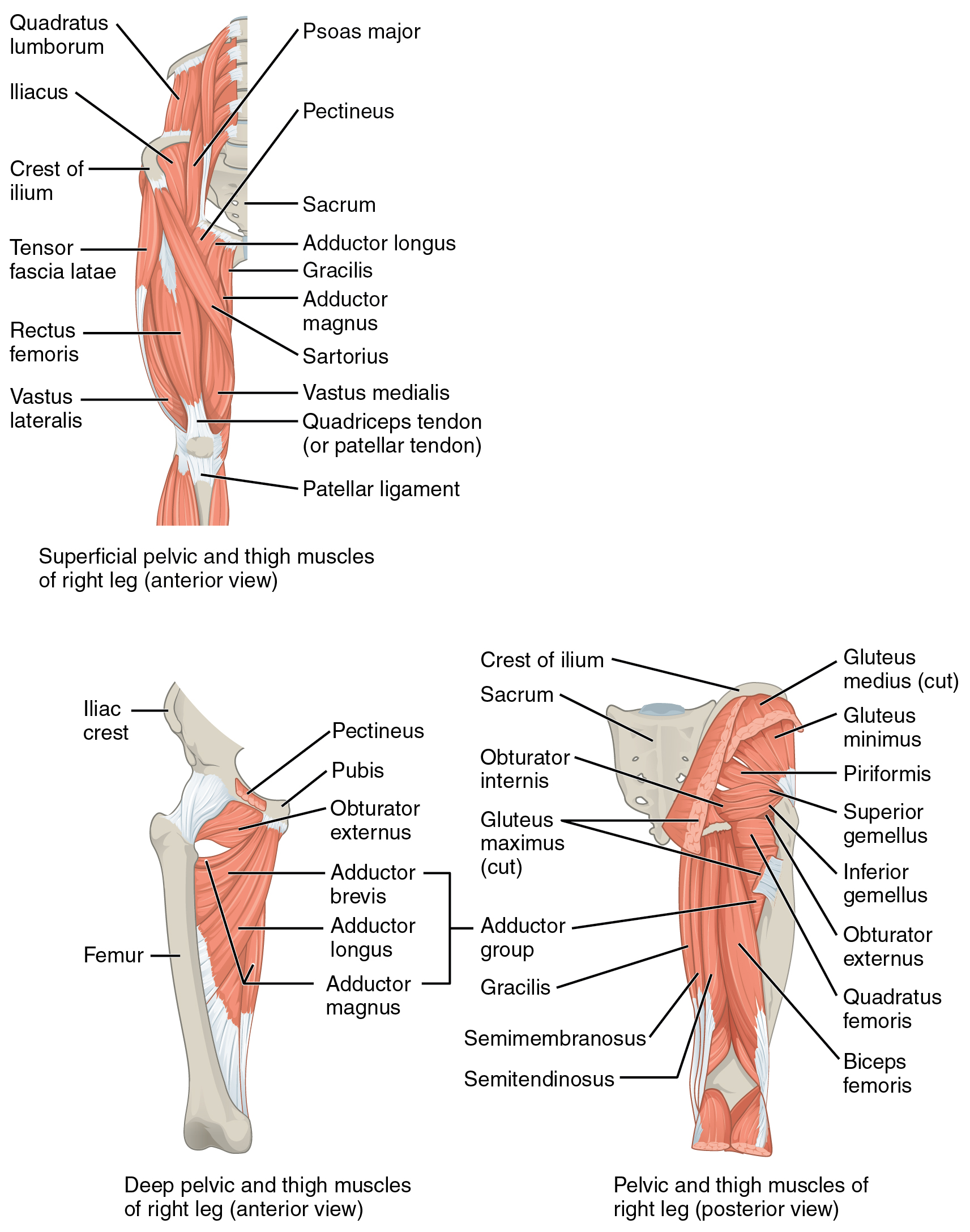
| Vastus medialis | Linea aspera Intertrochanteric line |
Tibial tuberosity via Patellar ligament Patellar retinacula |
Femoral n. L2 - L4 |
Knee: Extension |
Origin
Some sources suggest that the vastus medialis originates from the lateral lip of the gluteal tuberosity as well5.
Insertion
Nerve
Femoral nerve (L2, L3, L4)4
Action
Knee: Extension4
It should be noted that the vastus medialis is stronger and descends further than the vastus lateralis5.
VMO: Oblique Fibers
“The oblique fibers of the vastus medialis (frequently abbreviated as VMO) appear to be uniquely oriented to help offset at least part of the lateral pull exerted on the patella by the quadriceps muscle as a whole (review Fig. 13.24).106,195,287,289 Selectively cutting fibers of the VMO in cadaver specimens produces an average 27% loss in medial patellar stability across the tested knee range of motion.8 This finding may be difficult to apply to clinical settings because it is extremely rare for a person to have isolated paralysis of the VMO. For decades, however, anecdotal evidence has suggested a preferential atrophy in the VMO in persons with chronic patellofemoral pain or history of chronic dislocation, apparently from disuse or neurogenic inhibition. Recent data, however, question or refute the notion that the observed atrophy of the VMO is any more extreme than the atrophy often observed across the entire quadriceps muscle.102,103 Research that clearly supports the contention that abnormal neuromuscular function of the VMO is a cause of patellofemoral pain syndrome or chronic lateral dislocation is mixed and generally lacking.177 Nevertheless, the suspicion of preferential atrophy, inhibition, or delayed activation of the VMO has led to the development of multiple treatment approaches designed to selectively recruit, strengthen, or otherwise augment the action of this portion of the quadriceps. Although the rationale for this treatment approach is biomechanically sound, the ability to selectively alter the control, activation timing, or strength of only one component of the quadriceps remains a controversial topic.*”6
“The VMO arises from the adductor magnus tendon. The insertion site of the normal VMO is the medial border of the patella, approximately onethird to one-half of the way down from the proximal pole. If the VMO remains proximal to the proximal pole of the patella and does not reach the patella, there is an increased potential for malalignment.8”7
“The vector of the VMO is medially directed, and it forms an angle of 50–55 degrees with the mechanical axis of the leg. This oblique alignment provides a mechanical advantage for stabilizing the patella, which counterbalances the larger crosssectional area and thus the force-producing capacity of the VL.6 The VMO is least active in the fully extended position and plays little role in extending the knee, acting instead to centralize the patella within the trochlea and enhancing the efficiency of the VL.6 It is active in this function throughout the whole range of extension.”7
“The vastus medialis (Fig. 19-7) is composed of two parts that are anatomically distinct: the vastus medialis obliquus (VMO) and the vastus medialis proper or longus (VML). While there is some conjecture as to whether these are separate entities, most authors agree that the two components have differing functions due to their fiber orientation, attachments, and thus angle of force on the patella.6,29 The VML appears to have little biomechanical significance unlike its counterpart, the VMO.”7
“The vastus medialis consists of fibers that form two distinct fiber directions. The more distal oblique fibers (the vastus medialis “obliquus”) approach the patella at 50 to 55 degrees medial to the quadriceps tendon (see Fig. 13.24). The remaining more longitudinal fibers (the vastus medialis “longus”) approach the patella at 15 to 18 degrees medial to the quadriceps tendon.189 The oblique fibers of the vastus medialis extend farther distally than other muscular components of the quadriceps. Although the oblique fibers account for only 30% of the cross-sectional area of the entire vastus medialis muscle,267 the oblique pull on the patella has important implications for the stabilization and orientation of the patella as it slides (tracks) through the trochlear groove of the femur.”6
Vastus Medialis Lateralis (VML)
“The VML originates from the medial aspect of the upper femur and inserts anteriorly into the quadriceps tendon, giving it a line of action of approximately 15–17 degrees off the long axis of the femur in the frontal plane.”7
Knee Extensor mechanism
“Together, the quadriceps muscle, patella, and patellar tendon are referred to as the knee extensor mechanism”6
Rehabilitation
VMO Rehabilitation
“The VMO, which is frequently innervated independently from the rest of the quadriceps by a separate branch of the femoral nerve, is the first muscle of the quadriceps group to atrophy and the last to rehabilitate”8

