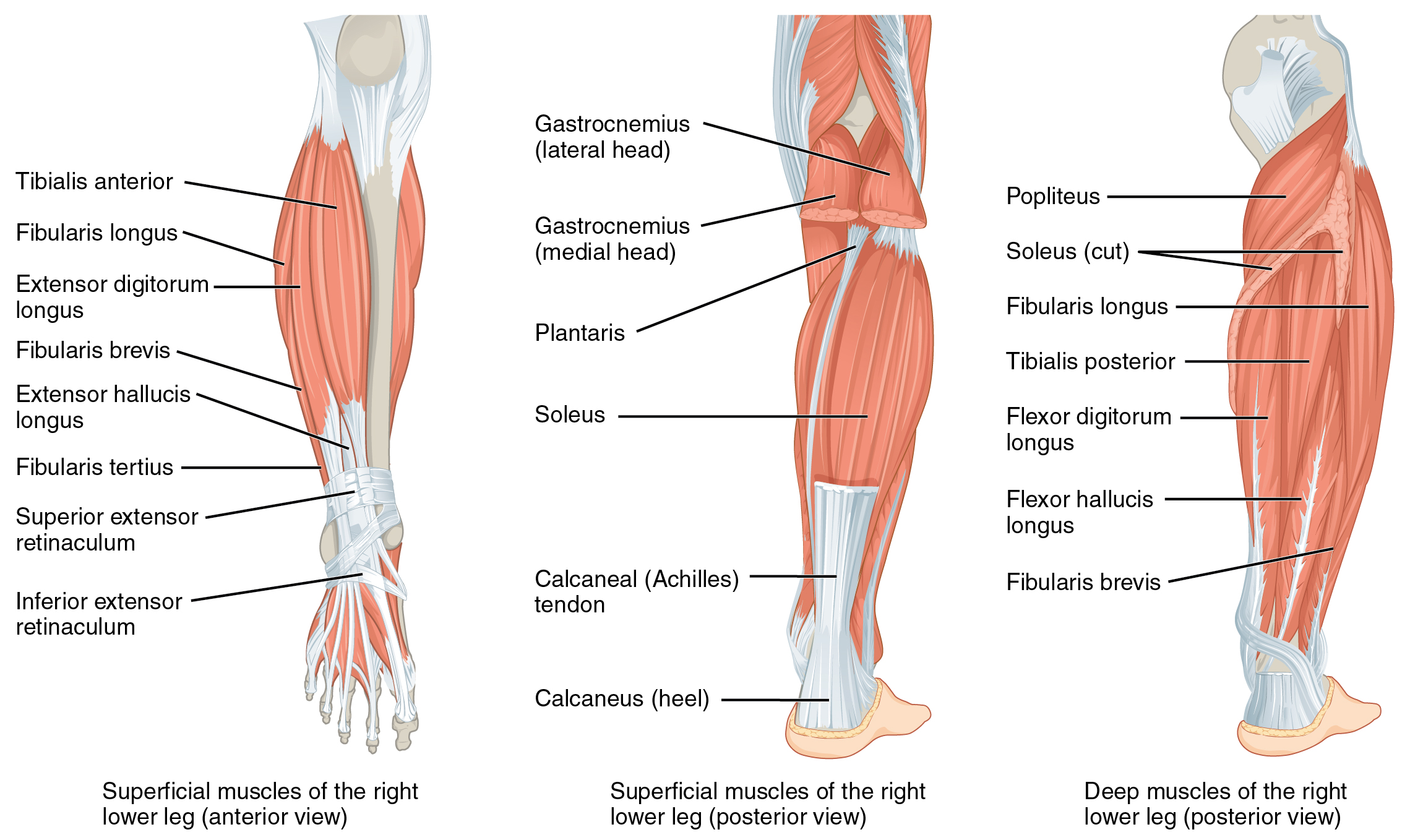
Tibialis Posterior Muscle

| Muscle | Origin | Insertion | Innervation | Action |
|---|---|---|---|---|
| Tibialis posterior | IO membrane Adjacent Tibia Adjacent Fibula |
Cuneiforms 2-4 MT bases |
Tibial n. L4 - L5 |
TCJ: PF Arch support: Transverse arch, Longitudinal arch |
Origin
Insertion
Nerve
Action
- TCJ: Plantarflexion6
- STJ: Inversion (supination)6
- Longitudinal Arch: Support6
- Transverse Arch: Support6
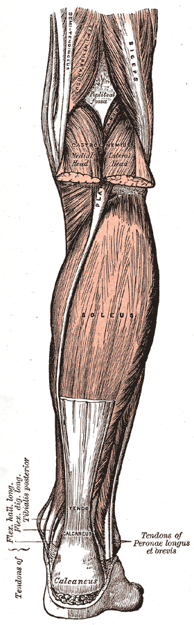
The posterior tibialis and flexor hallucis longus also improves the stability of ankle joint by creating a dynamic pinch between the malleoli, which improves joint contact during plantarflexio7.
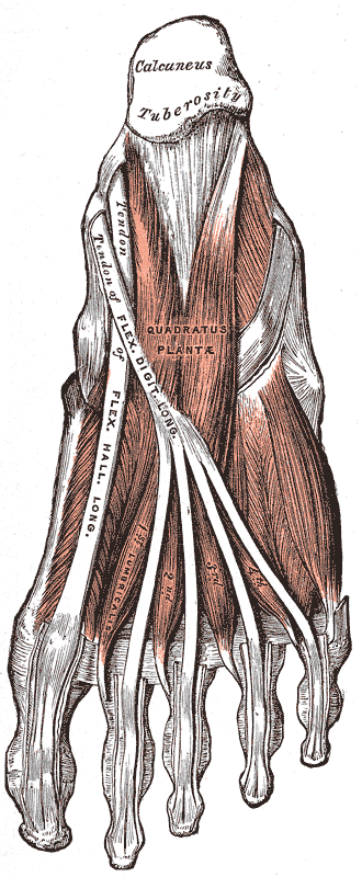
Tendon
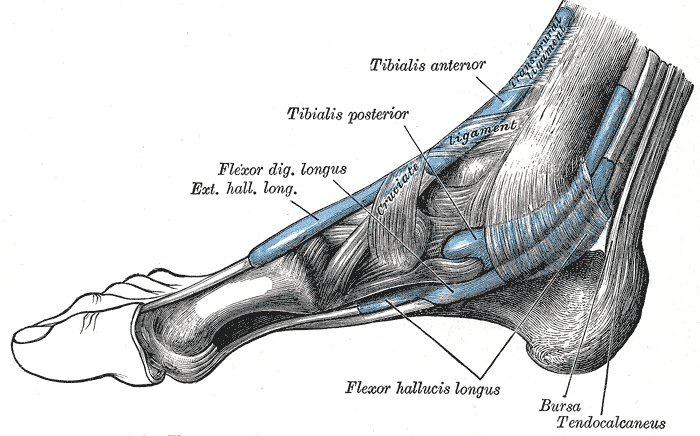
“This tendon is palpable at the level of the medial malleolus, especially with the foot held in plantar flexion and supination. Distal and medial to this tendon, the crossing of the FDL and flexor hallucis tendons can be felt.”8
Examination
Strength Testing
“The tibialis posterior muscle produces the motion of inversion in a plantar flexed position. The leg is stabilized in the anatomic position, with the ankle in slight plantar flexion. The plantar flexion is important to minimize the influence of the tibialis anterior muscle.77 Resistance is applied to the medial border of the forefoot into eversion and dorsiflexion (Fig. 21-24) VIDEO. The standing heel raise test can also be used to detect tibialis posterior weakness. It is thought that during a standing heel rise that the tibialis posterior and fibularis muscles co-contract to control hindfoot position. Thus, when the hindfoot everts during the heel-rise task, this is seen as a clinical sign of tibialis posterior weakness.”8
Dysfunction
Myofascial release
Pails & Rails
P.A.I.L.’s
- Plantarflexion
- Inversion
R.A.I.L.’s
- Extension
- Eversion
Stretch
Shin splints
Tibialis posterior dysfunction can lead to shin splints, more specifically posterior shin splints.