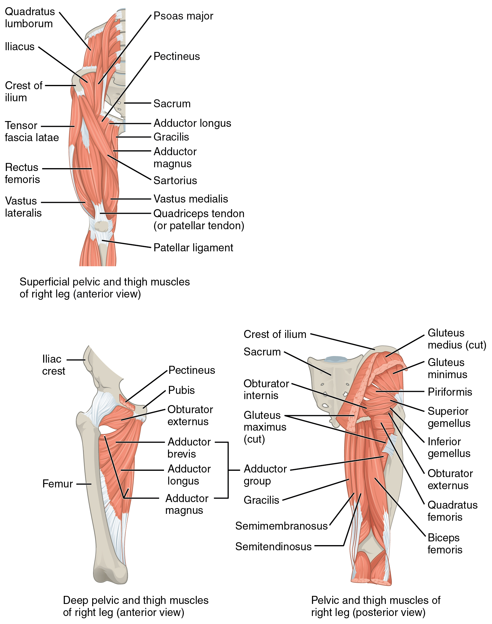
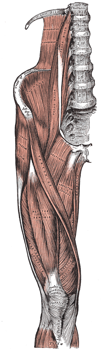
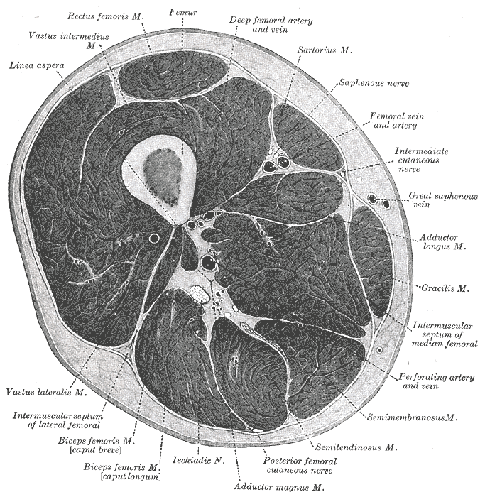
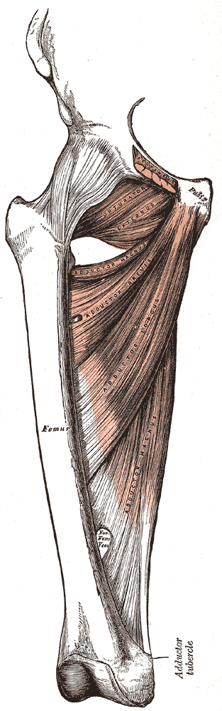
Adductor Magnus
| Muscle | Origin | Insertion | Innervation | Action |
|---|---|---|---|---|
| Adductor Magnus | Inferior pubic ramus Ischial ramus Ischial tuberosity |
Medial lip of Linea aspera Adductor tubercle |
Obturator n. Tibial n. L2 - L4 |
Hip: Adduction, Flexion, Extension, IR Pelvis: Frontal stabilization, Sagittal stabilization |
Origin
Insertion
Innervation
Action
- Hip: Adduction, Extension, and slight flexion (the tendinous insertion is also active in internal rotation)6
- Pelvic Stabilization: Coronal and Sagittal planes6
According to Brad Jones, the adductor magnus is a hip extensor until -20° Hip extension7.
Anatomy
“The anterior head of the adductor magnus has two sets of fibers: horizontal and oblique. The relatively small (and often poorly defined) set of horizontally directed fibers crosses from the inferior pubic ramus to the extreme proximal end of the linea aspera, often called the adductor minimus. The larger obliquely directed fibers run from the ischial ramus to nearly the entire length of the linea aspera, as far distally as the medial supracondylar line.”4
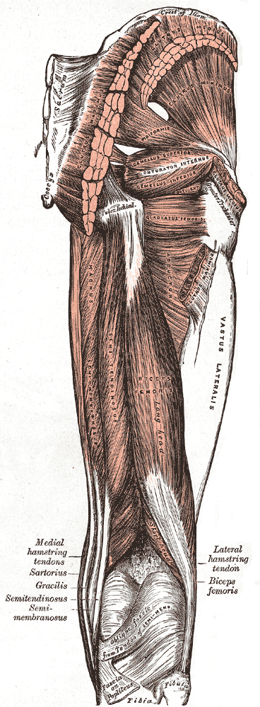
“The posterior head of the adductor magnus consists of a thick mass of the fibers arising from the region of the pelvis adjacent to the ischial tuberosity. From this posterior attachment, the fibers run vertically and attach as a tendon on the adductor tubercle on the medial side of the distal femur. The posterior head of the adductor magnus is innervated by the tibial branch of the sciatic nerve, as are most of the hamstring muscles. Because location, innervation, and action are similar to those of the hamstring muscles, the posterior head may also be referred to as the extensor head of the adductor magnus”4
Overview
“the adductor magnus is the largest of the adductor muscles, accounting for 60% of the total cross sectional area of the entire adductor muscle group.222 As a whole, the adductor magnus attaches proximally to the pelvis from two heads: an anterior head from the ischial ramus and a posterior head from the ischial tuberosity. Realize, however, that other anatomic classifications have been suggested”4
Palpation
See how to palpate this muscle along with other adductor muscles here.