| Muscle | Origin | Insertion | Innervation | Action |
|---|---|---|---|---|
| Splenius capitis | Nuchal lig. C7-T4 SP |
Lateral third of nuchal line Mastoid process |
Spinal nn. Posterior rami (lateral br.) C1 - C6 |
Bilateral: Head and neck extension Unilateral: I/L sidebend, I/L rotation |
Splenius Capitis


Origin
- Nuchal ligament4
- C7-T4 SP4
- Finando suggests that this muscle actually originates from the fascia associated with C4-T4 spinous processes3.
Insertion
The insertion is also called the “proximal attachment”3. The splenius capitis inserts proximally on:
The muscle runs deep to the sternocleidomastoid
Innervation
spinal nn. C1-C6 (Post rami, lateral branches)4
Action
Note
The splenius capitis is a much more dominant axial rotator than the splenius cervicis5
Clinical significance
Control of upright posture is possible through constant interaction between the visual and vestibular system with short range rotators including obliquus capitis posterior inferior, rectus capitis posterior major, splenius capitis, and SCM6.
Muscle groups
Splenius Capitis is part of the Superficial intrinsic back muscle group
Palpation
To locate the splenius capitis first palpate these structures:
To palpate splenius capitis:
- Have the patient in supine or seated with his/her back against the chair.
- Locate the muscular triangle between the SCM (anteriorly), upper trapezius (posteriorly), and the levator scapulae (distally).
- You should be able to palpate the taut bands of splenius capitis just proximal to the levator scapulae
Trigger point
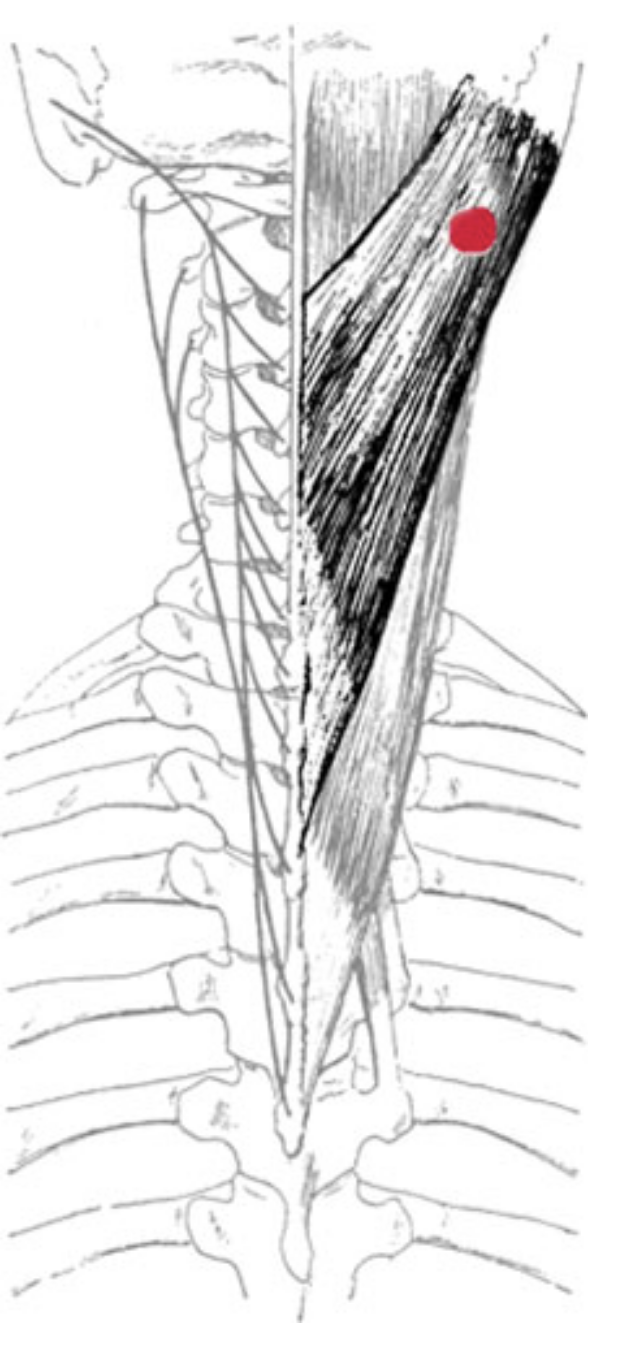
Pain pattern
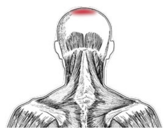
Etiology
Satellite Trigger Points
Associated zones, meridians, and points
- Dorsal zone
- Foot Tai Yang Bladder meridian
Stretch Exercises
Note
This stretch should be slightly more forward than lateral3
Clinical significance
1.
Betts JG, Blaker W. Openstax Anatomy and Physiology. 2nd ed. OpenStax; 2022. https://openstax.org/details/books/anatomy-and-physiology-2e/?Book%20details
2.
Donnelly JM, Simons DG, eds. Travell, Simons & Simons’ Myofascial Pain and Dysfunction: The Trigger Point Manual. Third edition. Wolters Kluwer Health; 2019.
3.
Finando D, Finando SJ, Finando D. Trigger Point Therapy for Myofascial Pain: The Practice of Informed Touch. Healing Arts Press; 2005.
4.
Gilroy AM, MacPherson BR, Wikenheiser JC, Voll MM, Wesker K, Schünke M, eds. Atlas of Anatomy. 4th ed. Thieme; 2020.
5.
Neumann DA, Kelly ER, Kiefer CL, Martens K, Grosz CM. Kinesiology of the Musculoskeletal System: Foundations for Rehabilitation. 3rd ed. Elsevier; 2017.
6.
Dutton M. Dutton’s Orthopaedic Examination, Evaluation, and Intervention. 5th ed. McGraw Hill Education; 2020.
Citation
For attribution, please cite this work as:
Yomogida N, Kerstein C. Splenius Capitis. https://yomokerst.com/The
Archive/Anatomy/Skeletal Muscles/Muscles of the back/Superficial
Intrinsic Back Muscles/Splenius capitis
muscle/splenius_capitis.html