Obturator Externus
| Muscle | Origin | Insertion | Innervation | Action |
|---|---|---|---|---|
| Obturator externus | Outer surface of Obturator membrane Adjacent bony boundaries |
Trochanteric Fossa | Obturator n. L3 - L4 |
Hip: Adduction, ER Pelvis: Sagittal Stabilization |
Origin
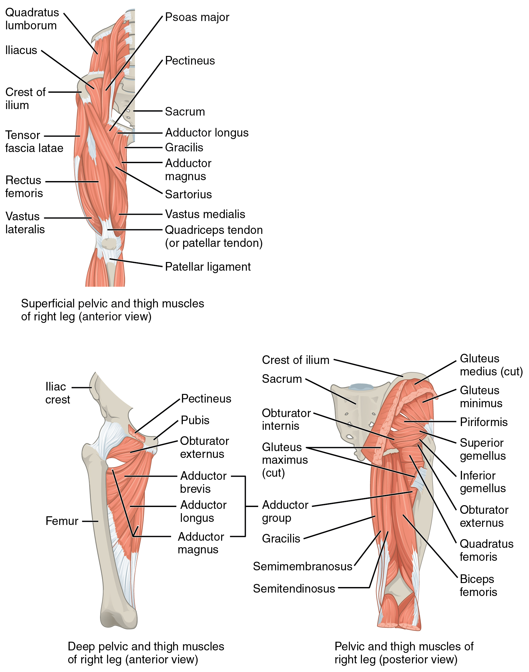
Outer surface of the obturator membrane and its bony boundaries5.
Path
From its origin the OE and its tendon wind posteriorly below the hip joint and run superiorly to its insertion on the floor of the trochanteric fossa6.
Insertion
Trochanteric fossa of the femur5
Innervation
Obturator N. (L3, L4)5
Action
According to Brad Jones6, the OE is an external rotator when in hip flexion, and acts to slightly flex the hip6.
At ~30-40° of internal rotation, the OE’s runs exactly inferior to the center of the joint, and thus stops functioning as an external rotator6. When the hip is internally rotated past 40°, the OE generates an external rotation torque6.
Overview
“The obturator externus muscle arises from the external side of the obturator membrane and adjacent ilium (see Fig. 12.14). The belly of this muscle is visible from the anterior side of the pelvis after removal of the adductor longus and pectineus muscles (see Fig. 12.26, left side). The muscle attaches posteriorly on the femur at the trochanteric fossa (see Fig. 12.6). (Based on its leverage to produce adduction, location, and innervation, the obturator externus is more anatomically associated with the adductor group of muscles than with the other five short external rotators. The obturator externus is innervated by nerve roots that originate from the lumbar plexus [via the obturator nerve], as are most of the other adductor muscles. The other small external rotators, in contrast, are innervated through the sacral plexus, with nerve roots as low as S2.)”7