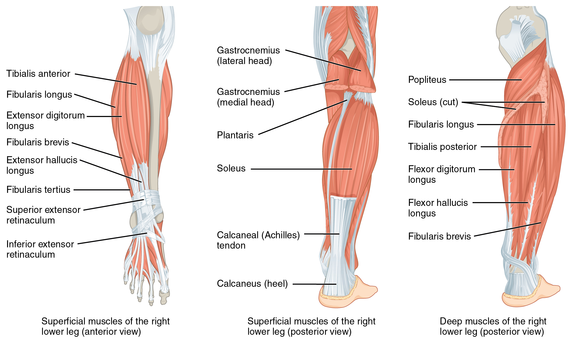
Fibularis Tertius
| Muscle | Origin | Insertion | Innervation | Action |
|---|---|---|---|---|
| Fibularis tertius | Anterior border of Distal Fibula | Shaft or Base of 5th MT | Deep Fibular n. L4 - L5 |
TCJ: DF STJ: Eversion |
Overview
Origin
The Fibularis Tertius generally originates from the distal fibula, either from the distal half (67%)6 or the distal third (22%)6. The other 11% of legs have an absent FT muscle belly, thus the tendon originates from the tendon of the EDL6.
Olewnik’s study was performed on Caucasian cadavers from a Polish population, thus may not be generalizable to other populations6
Insertion
Muscular Insetion
| Type | Prevalence | Attachment |
|---|---|---|
| I6 | 45%6 | Insertion on the shaft of the 5th MTP6 |
“Type I—single distal attachment. The tendon inserts into the shaft of the fifth metatarsal bone. This type was found in 41 lower limbs (45%)—Figure 2a.”6
“Type II—single distal attachment. The tendon is characterized by a very wide insertion into the base of the fifth metatarsal bone. This type was found in 20 lower limbs (22%)—Figure 2b.”
“Type III—single distal attachment. The tendon is characterized by a very wide insertion into the base of the fifth metatarsal bone, the base and shaft of the sixth metatarsal bone, and the fascia covering the fourth interosseous space. This type was found in 15 cases (16.5%)—Figure 2c.”
“Type IV—bifurcated distal attachment. The main tendon inserts into the base of the fifth metatarsal bone, and the accessory band inserts into its shaft. This type was observed in eight lower limbs (8.8%)—Figure 3a.”
“Type V—bifurcated distal attachment. The main tendon is characterized by a very wide insertion into the base of the fifth metatarsal bone, and the accessory band inserts into the base of the four metatarsal bone. This type was observed in five lower limbs (5.5%)—Figure 3b.”
“Type VI—this is characterized by fusion with an additional band of the fibularis brevis tendon. This fusion gives rise to the fourth interosseus dorsalis muscle. Type VI was observed in two cases (2.2%)—Figure 3c.”
Middle shaft or base of 5th MT6
Innervation
Deep Fibular N. (L4, L5)7
Action
Function
Gait
EMG studies indicate that Fibularis Tertius works in conjunction with EDL during the swing phase of walking to create dorsiflexion and eversion and to elevate the foot and toes from the ground6.
Eversion
Fibularis teritus, Fibularis Longus, and Fibularis Brevis work together to perform Eversion and prevent hyper-inversion6.


