Tone Management
Spasticity and Rigidity
What is Muscle Tone
“Tone” refers to a relaxed muscle’s resistance to passive stretch/tension
The muscle resists the stretch through both active and passive mechanisms
Passive component
The passive mechanisms refer to viscoelastic structures that are non-contractile.
- Actin & Myosin
- Connective tissue
- etc
Forces created through passive mechanisms are invisible to EMGs
Active component
The active mechanisms refer to contractile motor units
These are controlled by spinal and supraspinal mechanisms
Forces created through active mechanisms can be measured by EMGs
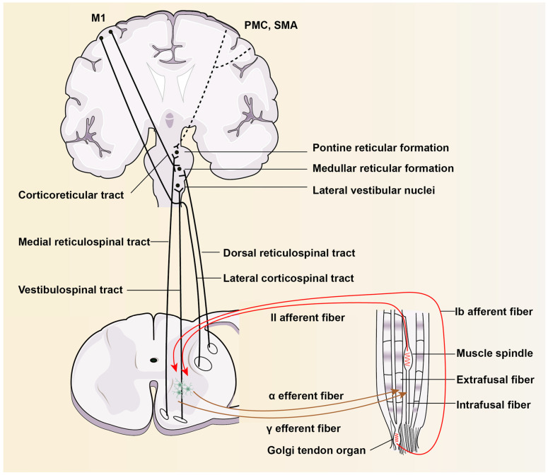
Anatomy
- Muscle spindle: Intrafusal fibers
- Muscle belly: Extrafusal fibers
- Tendon: Connective tissue connecting muscle to bone
Intrafusal Fibers
- “Muscle spindle”
- Sensory organs
- Detects changes in Length AND Velocit
Extrafusal Fibers
- “Muscle belly”
- Contraction creates force and movement
Tendon
- Connects muscle to bone
Intrafusal fibers
Two types: Nuclear bag fibers and Nuclear chain fibers
Nuclear bag fibers:
- Length + velocity
- Ia Afferents → SC
Nuclear chain fibers:
- length
- Ia and II Afferents →SC
Both Synapse onto Gamma motor neurons in Spinal Cord
Extrafusal fibers
- Contracts muscle itself
- Activated by Alpha Motor Neurons from SC
Stretch Reflexes
Muscle spindles generate tone via the stretch reflex (dynamic or static):
2 types
Dynamic: sudden rapid stretch of a muscle → Ia → SC→ alpha motor neuron causes sudden contraction of muscle
Static: Sustained stretch→ type II → spinal cord → alpha motor efferents → cause asynchronous contraction of muscle fibers (motor units not all discharging together) = mild sustained contraction of these fibers as long as it is stretched.
This is the physiological basis of maintaining muscle tone
Mechanism
Muscle Stretches → Sensory info send to spinal cord → Muscle contracts to prevent overstretch damage
- Muscle stretches
- Intrafusal fibers sense stretch
- Ia + II afferents send signals to SC
- Synapses onto agonist Alpha motor efferents
- Agonist muscle contracts
Alpha motor neurons → Extrafusal fiber contracts to prevent overstretching Quadriceps contract
Reciprocal Inhibition
Within spinal cord, Ia & II afferents also synapse onto:
- Inhibitory Interneurons of alpha motor neurons
- Inhibits alpha motor neurons of antagonist muscles while agonist contracts
- Result: Antagonist muscle cannot contract preventing further stretch of the agonist
How Do They work during muscle stretch
Patellar Tendon Reflex
- Hammer hit at tendon
- Quadricep muscle is stretched → Stretching both the intrafusal and extrafusal fibers
- Intrafusal fibers detect stretch (length/velocity)
- Type Ia and II afferent fibers
- Signals carried to DRG
- To dorsal horn of spinal cord and synapses on:
From dorsal horn to α-efferent
- Motor neuron (alpha/efferent)
- quad extrafusal fiber and cause it to contract to protect the muscle from overstretching (a monosynaptic reflex)
From dorsal horn to interneuron
- Interneuron → reciprocal inhibition
- Reciprocal inhibition
- inhibits alpha-motor efferents of antagonist muscles (Hamstrings relax)
Recurrent inhibition
- Motor neuron fires → activates inhibitory interneuron
- Inhibits the same neuron or a group of neurons.
- Mediated by Renshaw cells in spinal cord
This feedback loop helps regulate motor neuron activity, preventing excessive firing and contributing to the fine-tuning of motor control.
Found to be increased in those with SCI - Overexcitation1.
VERY DEBATED IF INVOLVED IN SPASTICITY
Alpha-Gamma Coactivation
Body needs to maintain stretch sensation when a muscle is contracting:
- Muscle contraction →
- Extrafusal fibers AND intrafuMuscle contraction → sal fibers contract→
- Muscle spindle tension is maintained due to the intrafusal fiber contraction →
- Maintaining the firing rate for the type Ia fibers and keeps it in an optimal range for detecting a change in muscle length
HYPOTHETICALLY if ONLY alpha-motor efferents are excited and Extrafusal fibers contract → reduces tension on muscle spindle → desensitization → decreased stretch proprioception info
HOWEVER this is not exactly what happens because the body needs to have stretch sensation during contraction/shortening!
Intrafusal fibers
- Muscle stretches
- Intrafusal fibers sense stretch
- Ia + II afferents send signals to SC
- Synapses onto agonist Alpha motor efferents
- Synapses onto inhibitory interneurons
- Synapse on Agonist gamma-motor efferent
- Stimulates agonist intrafusal fiber contraction → tensioning muscle spindle to maintain stretch sensitization
- Synapse on Inhibitory Interneuron of antagonist gamma-motor efferent
- Inhibits gamma motor neuron to antagonist muscle, decreasing tension on antagonist muscle spindle (desensitized to stretch)
Intrafusal and Extrafusal fibers’ role in proprioception
To provide proprioception, the intrafusal fibers must contract and stretch in parallel with the extrafusal fibers MM Stretch → both intrafusal and extrafusal are stretched → type Ia/II → sensitive to stretch During a stretch, the stretch on the intrafusal fibers provide proprioceptive information of a muscle stretch MM Contraction → you would expect extrafusal contraction and intrafusal fibers to not contract However, not exactly the case;alpha gamma co-activation Alpha and gamma motor neurons contract in parallel in order to maintain tension on the intrafusal sensory fibers to provide proprioception during contraction
GTOS: Prevents tendon avulsion
Golgi tendon organs (GTOs) are sensory organs found within tendons
These organs detect tendon stretch and send sensory signals via Type 1b afferents → spinal cord.
GTO Example: Ego bicep curling
Muscle contraction → tendon lengthens → GTOs are proprioceptive/sense length → type 1b afferents → SC → DRG → synapses onto 2 interneurons:
Inhibitory interneuron
The inhibitory interneuron is activated which inhibits the alpha motor neuron to the bicep → Decreasing tension of agonist (bicep)
Excitatory Interneuron
Antagonist contraction to pull forearm in opposite direction Interneuron → alpha motor neuron → Excites antagonist (triceps)
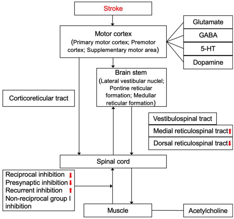
Supraspinal influences
CST and RST tracts
Corticospinal Tract
- Cerebral cortex → crosses at pyramidal decussation → motor neurons
- Inhibits lower motor neurons
- Gives off corticoreticular pathway on way down → medullary reticulospinal nuclei → reticulospinal (RST) tract
- Inhibits lower motor neurons
slide 19
- Supraspinal: facilitatory and inhibitory long tracts + cerebellum,BG, limbic system and more thought to modulate muscle tone…
- Inhibitory: All work to reduce excitability of motor neurons/dec muscle tone:
- CST: inhibits spinal reflexes, helping with fine tuning of motor commands, suppressing excessive reflex activity. Crucial for voluntary motor control
- Dorsal RST: suppresses muscle tone, usually during REM sleep or motor control tasks
- Facilitatory: All work to increase excitability of motor neurons, increasing tone
- VST: tonic drive reflects motion and position of head and projects to extensor muscles, provides drive to facilitate antigravity support. Disruption leads to dec’d extensor tone/postural instability
- Medial RST: excites motor neurons controlling axial/proximal limb muscles, supporting postural adjustments/locomotion
Hypertonia/Hyperreflexia in UMN Lesions
Where does it all go wrong?
UMN Lesion → Hypertonia/Reflexia
Damage in Cerebrum:
- Inhibition of medullary RST/CST → Disinhibition of motor neurons:
- Increase alpha motor excitation → inc in extrafusal fiber contraction → HYPERTONIA
- Increase gamma motor excitation → inc in intrafusal fiber contraction → sensitized intrafusal fibers → reflex pathway activates → HYPERREFLEXIA
slide 22
MANY pathways are involved… medullary RST has a large effect on Hypertonia/Hyperreflexia
Studies show later on RST may take over more muscle mvmt to make up for dysfunctional CST in Spasticity (Sangari et al, 2019)
Clonus
Reciprocal inhibition (which relaxes the antagonist muscle normally) is reduced with supraspinal lesions, leading to contract-relax cycles as muscles are stretched/contracted.
Spasms
This heightened reflex activity can result in involuntary muscle spasms, which are sudden, often painful contractions of the muscle in response to stretch
Spasticity Versus Rigidity
| Differentiating Points | Spasticity | Rigidity |
|---|---|---|
| Velocity dependency | Yes | No |
| Resistance to movement | In one direction (flexion or extension) | In both directions |
| Length dependency | Yes | No |
| Type of hypertonicity | Clasp-knife | Lead pipe or Cog-wheel |
Clasp Knife Effect
- High velocity of quick stretch activates nuclear bag then 1a afferents → sudden contraction
- Further into the stretch, the stretch is detected further by nuclear bag and nuclear chain → 1a and II afferents activate → alpha motor neuron → contract → maintains the resistance through some of the range
- Release: Golgi tendon organ (Ib afferent) activate, which inhibits α-motor neurons, reducing resistance, allowing some stretch. → like a tardieu 2 - catch + release
| Grade | Criteria |
|---|---|
| 0 | No resistance throughout the course of the passive movement |
| 1 | Slight resistance throughout the course of the passive movement, followed by release |
| 2 | Clear catch at precise angle, interrupting the passive movement, followed by release |
| 3 | Fatigable clonus <10 seconds when maintaining pressure occurring at precise angle |
| 4 | Infatigable clonus >10 seconds when maintaining pressure occurring at precise angle |
Why are only some muscles spastic?
- More Excitable To Begin With
- Biceps, PFs are more excitable/experience more spasticity because they are more used throughout our daily life
- Excitability amplified after injury
Synergies + Spasticity
Why do we have them?
- Error compensation - if one muscle is weak, others help achieve motion
- Energy efficiency- CNS reduces complexity of mvmt by organizing muscles into synergies, allows for quick/automatic mvmts without higher level control
- UE = flexor synergy dominant LE= extensor synergy dominant
Synergies
After UMN lesion
- CST is damaged, body increases reliance on RST - less capable of fine motor, better at gross motor control - leading to synergistic mvmts
- RST Is disinhibited (excited), causing involuntary co contraction – ie trying to lift shoulder, elbow + wrist flex too
MMTs are not indicated for Synergistic Muscles
Synergies make MMTs inaccurate
Co-activation of other muscles makes it impossible to isolate 1 muscle
Not purely volitional movement, but rather more automatic driven (RST)Mooney2024?
Benefits of Spasticity
Not always harmful!
- Can improve functional activities like transfers, standing, gripping
- Acute increases can also be a sign: UTI, fracture, pressure sore
- Improves venous return/circulation
- Protects against DVTs
- Reduces fracture risk, decreases risk of osteoporosis
- Protects against muscle atrophy
- Marker of motor recovery potential
Tonic & Phasic Spasticity
- Velocity dependent increase in tonic stretch reflex (tone), hyperexcitability of stretch reflex
- Spasticity can be “phasic” or “tonic” d/t predominant involvement of different components of stretch reflex.
- Phasic involuntary mm contraction after quick stretch, clonus. Hyperactive phasic stretch reflex
- Tonic (static): hyperactive tonic stretch reflex seen in chronic spasticity/non ambulatory pts, passive stretch at ankle (experts argue this is just rigidity/severe hypertonia rather than true spasticity)
- Happens after spasticity affects patient over time
| Feature | Phasic Spasticity | Tonic Spasticity |
|---|---|---|
| Trigger | Rapid stretch | Sustained posturing |
| Velocity-Dependent | Yes | No |
| Neural Mechanism | Exaggerated stretch reflex (Ia afferents) → activates alpha motor neurons | Continuous excitation of α- and γ-motor neurons from Ia and II afferents |
| Clinical Sign | Clonus, quick spasms | Constant stiffness, resistance to movement |
Pathophysiology
Spasticity is mostly due to dis-inhibition of the medullary (dorsal) Reticulospinal tract
- Spasticity may be maintained through facilitatory influences of the medial (pontine) RST
- Vestibulospinal tract plays a minor role
- As shown by sectioning studies of the human spinal cord in those w/ spasticity
- Other brain regions may also be involved: BG, cerebellum, etc
- Studies are still examining all influences on spasticity/tone – a lot is still unknown…
Spinal Influence/SCI spasticity
Spinal influence:
Many different ways to inc excitation or dec inhibition in spinal cord
Descending monoaminergic drive normally excites alpha motor neurons and inhibits dorsal horn/sensory input
SCI Chronicity
Acute stage of SCI: loss of monoaminergic influence → causes hypo excitability of motor neurons and excitation of sensory input
Chronic SCI Spasticity →Product of overactive motor neurons Weeks after disconnection from supraspinal input, motor neurons transition into an excitable state, really responsive to excitatory NTs like glutamate. Leads to muscle contractions, easily triggered by touch/mm stretching.
SCI SEVERITY
Incomplete SCI
Incomplete/partial myelopathy involving lateral funiculus: If there is involvement of CST only, it will result in weakness, hypotonia and loss of superficial reflexes. If there is additional involvement of dorsal RST, spasticity and hyperreflexia will develop due to unopposed activity of medial RST. Spasticity will be predominant in antigravity muscles and will result in paraplegia in extension and extensor spasms. Flexor spasms can occur if FRA are activated by pressure sores.
On the other hand, if there is involvement of dorsal RST only with sparing CST, there will be spasticity without much weakness.
Complete SCI
Complete myelopathy with involvement of all four tracts: Spasticity will be less in this case because of lack of facilitatory input from medial RST and VST. Disinhibition of FRA will result in paraplegia in flexion and flexor spasms.
Parkinsonian Rigidity
Rigidity: Increased resistance to passive movement, affecting both flexor and extensor muscles in Parkinson’s disease
Neural mechanisms are incompletely understood
Types of Rigidity
| Cogwheel | Leadpipe |
|---|---|
| Rigidity + tremor | Smooth, consistent |
| Tonic increase in muscle tonewrightAxialHypertonicityParkinsons2007? | EMG studies show phasic bursts of muscle activitymearaRelationshipElectromyographicActivity1992? |