flowchart BT

  mesencephalic_response[Modulating the bite]
  mesencephalic_response --> mesencephalic_nucleus[Mesencephalic Nucleus]
  proprioceptive_fibers[Proprioceptive fibers from extraocular muscles and masticatory muscles]

  teeth[Teeth] -- proprioception --> proprioceptive_fibers
  periodontium[Periodontium] -- proprioception --> proprioceptive_fibers
  hard_palate[Hard Palate] -- proprioception --> proprioceptive_fibers
  temporomandibular_joint_capsule[Temperomandibular Joint Capsule] -- proprioception --> proprioceptive_fibers

trigeminal_ganglion[Trigeminal Ganglion]


tactile_and_pressure_receptors[Receptors] -- Tactile & Pressure --> upper_face[Skin of Upper Face]
tactile_and_pressure_receptors -- Tactile & Pressure --> middle_face[Skin of Middle face]
tactile_and_pressure_receptors -- Tactile & Pressure --> lower_face[Skin of Lower Face]

upper_face --> opthalmic[Opthalmic Division V1]
middle_face --> maxillary[Maxillary Division V2]
lower_face --> mandibular[Mandibular Division V3]

opthalmic --> trigeminal_ganglion
maxillary --> trigeminal_ganglion
mandibular --> trigeminal_ganglion

trigeminal_ganglion -- Tactile & Pressure --> trigeminal_sensory_nucleus[Trigeminal Sensory Nucleus]

trigeminal_sensory_nucleus -- Tactile & Pressure --> VPM[Thalamus VPM]
CNV Trigeminal Nerve
 and the structures it innervates in the face and mouth.png)


Note
Overview
CN V Trigeminal nerve is the largest cranial nerve and provides general somatic sensory to the scalp, teeth, oral and nasal cavity as well as branchial motor functions to the muscles of mastication and some surrounding musculature3,5.
Additional functions
- Additionally, The trigeminal system plays a role in facial and dural sensitivity and has also been considered a component in brain nociceptive innervation6.
- “Proprioceptive nerve fibers from the masticatory muscles and (probably) extraocular muscle”5.
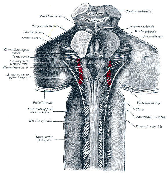
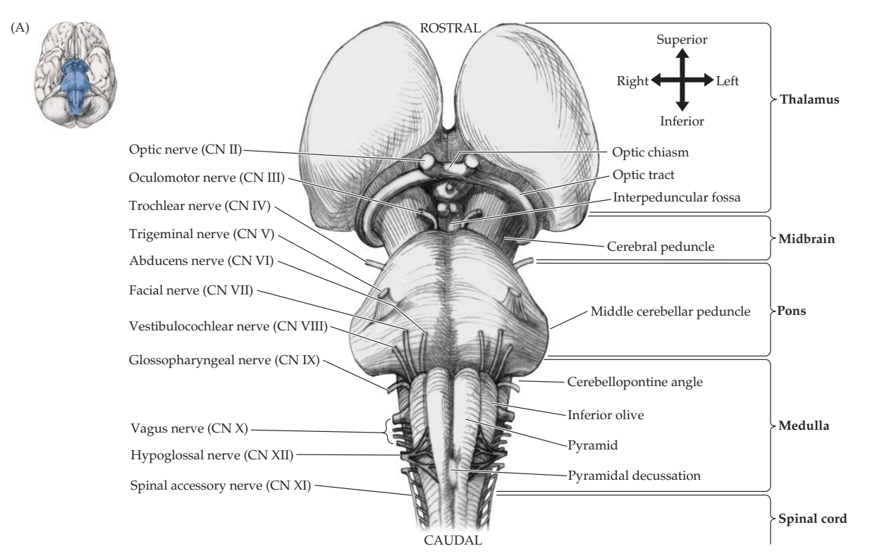
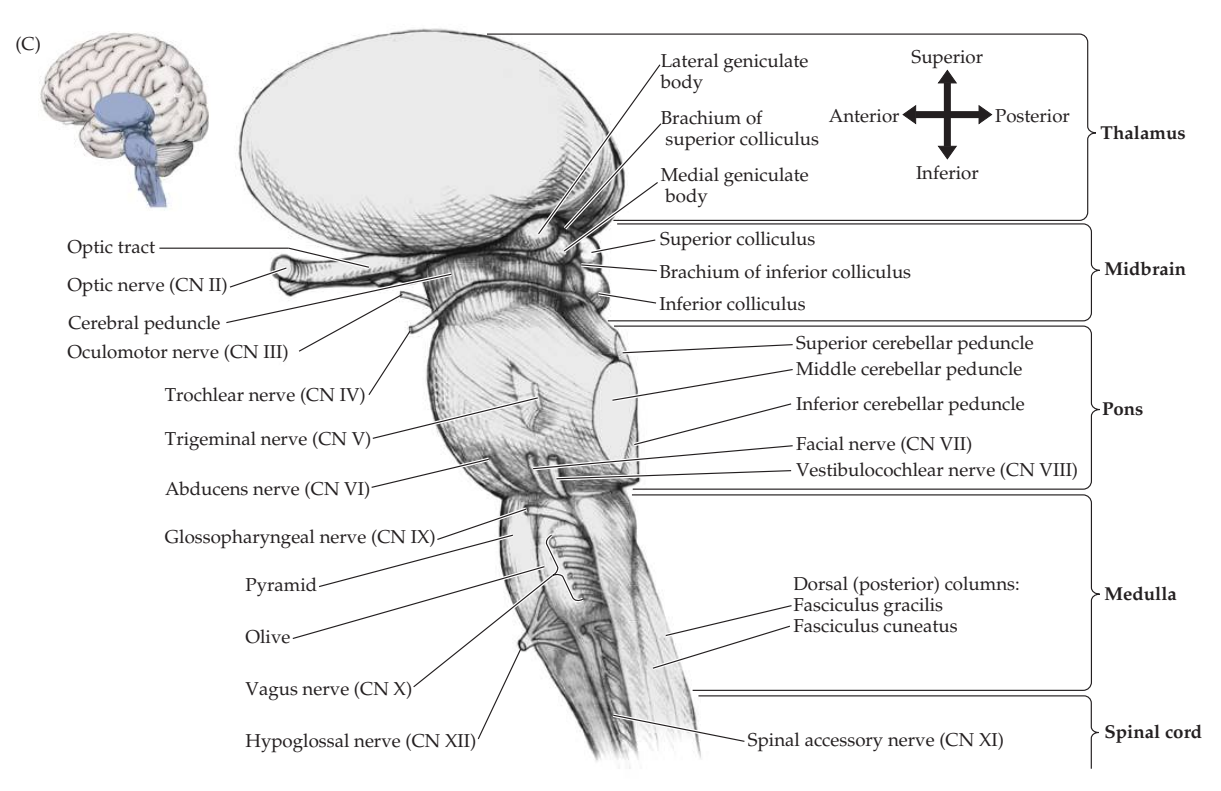
The Trigeminal nerve’s course can be broken down into: - Brainstem - Cisternal segment - Meckel’s cave segment - Trigeminal ganglion - Pperipheral divisions: Opthalmic division (V1), Maxillary division (V2), and Mandibular division (V3).
Why should I care about this nerve? - The trigeminal nerve is the anatomical substrate of several pathologies or conditions, including: Headaches (primary or secondary), trigeminal neuralgia, and alternate types of orofacial pains6. - Understanding the complex anatomical arrangement of CN V’s pathway is crucial to understand these conditions’ pathophysiology and treatment6.
Trigeminal Nuclei
There are 4 trigeminal nuclei (3 sensory and 1 motor):
- Mesencephalic nucleus (Conveys afferent proprioceptive fibers from extraocular and masticatory muscles and allows for bite modulation5)
- Trigeminal sensory nucleus
- Conveys touch and proprioception of the jaw area -ninjanerd
- Conveys tactile and pressure sense5
- Trigeminal motor nucleus (Modulates degree of bite)
- Spinal trigeminal nucleus Conveys touch, pain, pressure, and proprioception from the entire face
Anatomy
Brainstem
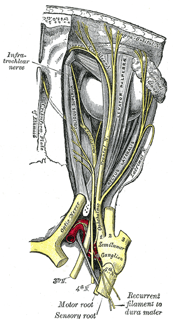
Trigeminal Root (Cisternal Segment)
- The 4 trigeminal nuclei (3 sensory, 1 motor) extend throughout most of the brain stem5
- “The trigeminal root is composed of the large sensory root and the small motor root”5
- “The sensory root receives somatosensory sensation from the entire face , temple, external acoustic meatus, and the anterior scalp as far posterior as the vertex of the skull”5
Note
The trigeminal nerve supplies somatosensation to the entire face except the angle of the jaw innervated by the cervical plexus5
The proprioceptive impulses from the masticatory muscle run through the motor root to enter the mesencephalic nucleus5
flowchart TD
Sensory Root (Portio Major)
Sensory Pathway
- Somatosensory afferent nerves convey crude touch, pain, and temperature sensation from the face and mouth to the lateral pons3.
- These nerve fibers then descend the spinal trigeminal tract to synapse in the spinal trigeminal nucleus3.
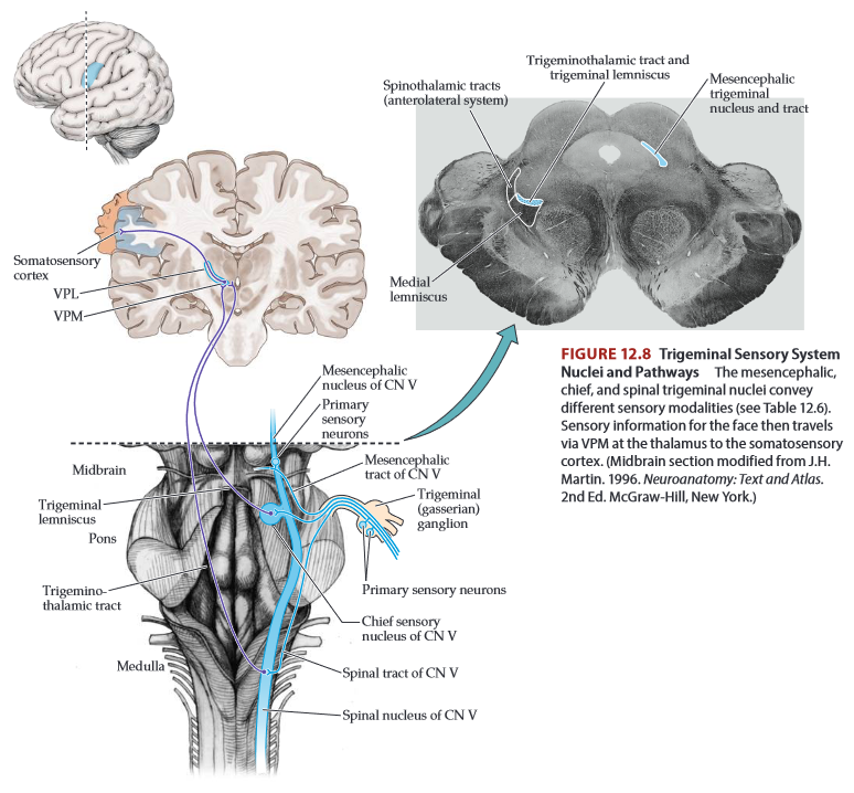
- . Similarly, the spinal trigeminal tract is analogous to Lissauer’s tract (see Figures 6.4 and 7.2). Secondary sensory neurons from the spinal trigeminal nucleus cross the brainstem to ascend as the trigeminothalamic tract (or ventral trigeminothalamic tract). The trigeminothalamic tract is analogous to the spinothalamic tract (see Table 12.6), and the pathways travel together to the thalamus (see Figures 12.8 and 14.3). Trigeminothalamic tract fibers synapse in the thalamic ventral posterior medial nucleus (VPM), and tertiary sensory neurons then travel in the internal capsule to the primary somatosensory cortex. Like the anterolateral systems in the spinal cord, there are also pathways from the spinal trigeminal nucleus to intralaminar thalamic nuclei, the reticular formation, and other areas, to mediate the affective and arousal aspects of facial pain.
In summary, it is generally believed that sensory fibers involved in the conduction of pain and temperature spread over the trigeminal sensory nucleus complex (TSNC) and then cross over to the contralateral thalamus and cerebral cortex7
Sensory Function
- V1 Opthalmic supplies the upper face
- V2 Maxillary supplies the middle face
- V3 Mandibular supplies the lower face
The trigeminal nerve also provides touch and pain sensation for the nasal sinuses, inside of the nose, mouth, and anterior two-thirds of the tongue.
Dysfunction
Motor Root (Portio Minor)
Motor Pathway
- The motor plan is sent from the cerebrum/cerebellum inferiorly to the trigeminal motor nucleus
- From there, the motor signal exits the trigeminal motor nucleus and passes anteriorly in the pons8
- The motor root emerges from the ant-lat aspect of the pons8
- The motor root is anterior and medial relative to sensory root8
- Next, the motor root passes through the posterior fossa and then through the dura mater below the attachment of the tentorium8
- The motor root then enters Meckel Cave8
- Upon its exodus from the skull, the motor root joins the sensory fibers in the mandibular (V3) division to form the mandibular nerve
- The mandibular nerve (V3) connects the motor root to the masticatory muscles (masseter, temporalis, and medial and lateral pterygoid muscles)8
- In addition, motor fibers are given off to the tensor tympani, tensor veli palatini, and mylohyoid muscles, and to the anterior belly of the digastric muscle”8
Motor Function
Meninges Sensitivity
The trigeminal system not only supports sensation to the face, but also the dura and pia6.
Brain innervation
Pathways
- The trigeminal nerve exits the brainstem from the ventrolateral pons3.
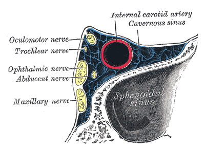
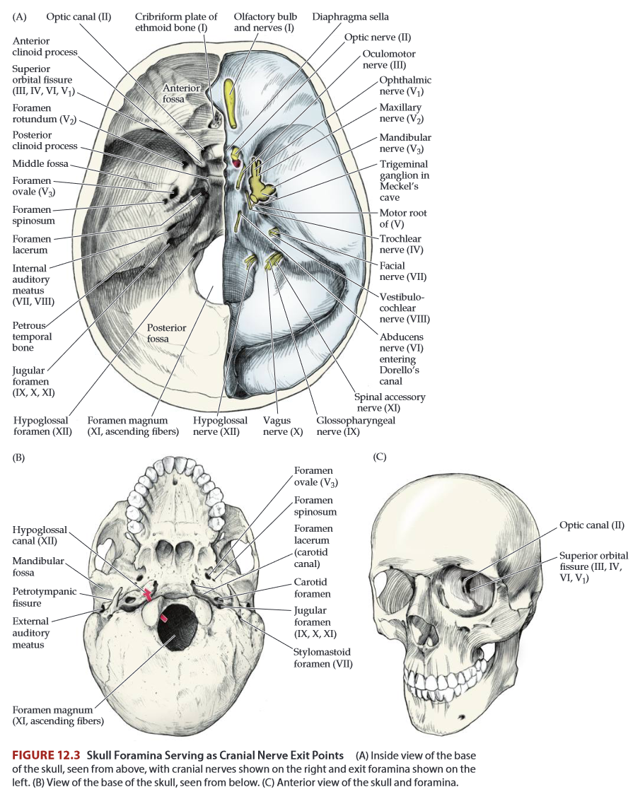
- Next CN V enters Meckel’s Cave (a small fossa posterior and inferolateral to the cavernous sinus)3.
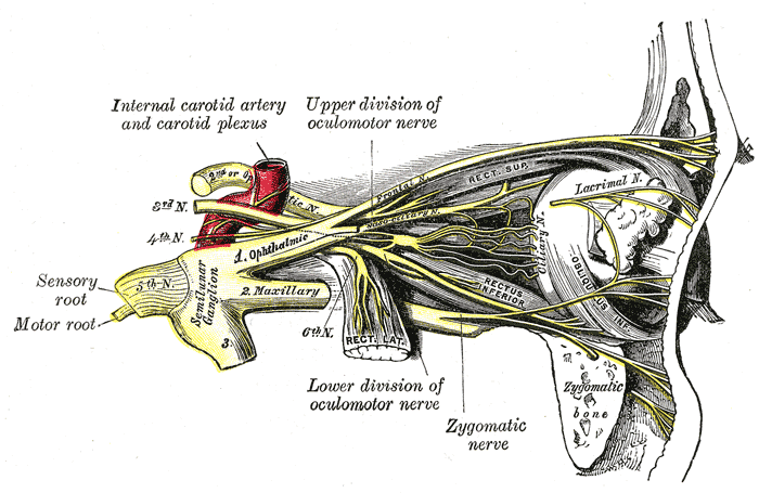
- CN V synapses on the trigeminal ganglion (sensory ganglion) in Meckel’s cave9.
- The ophthalmic division (V1) travels through an inferior section of the cavernous sinus and exits the skull through the superior orbital fissure3.
- The maxillary division (V2) exits via the foramen rotundum3
- The mandibular division (V3) via the foramen ovale3
Mnemonic
A way to remember the exit points of each trigeminal division is “Standing Room Occupancy”, or SRO (for Superior, Rotundum, Ovale)
In addition, pain sensation for the supratentorial dura mater is supplied by the trigeminal nerve, while the dura of the posterior fossa is innervated by CN X and upper cervical nerve roots.
Skin of face –> Receptor –> fine touch/dental pressure –> DIVISION –> Chief trigeminal sensory nucleus –> Trigeminal lemniscus –> VPM of thalamus
Skin of face –> Receptor –> Proprioception –> DIVISION –> Mesencephalic trigeminal nucleus –> ?
Skin of face –> Receptor –> Crude touch / Pain / Temperature –> DIVISION –> trigeminothalamic tract –> VPM of Thalamus
Trigeminal Ganglion
(AKA “semilunar ganglion” or “gasserian ganglion”)
| Nucleus | Sensory Modalities | Main pathway to thalamus | Main thalamic nucleus |
|---|---|---|---|
| Mesencephalic trigeminal nucleus | Proprioception | - | - |
| Chief trigeminal sensory nucleus | fine tough; dental pressure | Trigeminal Lemniscus | VPM |
| Spinal trigeminal nucleus | Crude touch; Pain; Temperature | Trigeminothalamic tract | VPM |
| Posterior column nuclei | Fine tough; proprioception | Medial lemniscus | VPL |
| Dorsal horn | Crude touch; Pain; Temperature | Spinothalamic Tract | VPL |
Trigeminal Neuralgia
Tic Doloureux
Testing
External Resources
- Ninja Nerd’s Trigeminal Nerve video
- Brazis’s localization in neurology contains a solid overview and really good DDX8
References
1.
Boundless. Boundless Anatomy and Physiology. Open Education Resource (OER) LibreTexts Project https://university.pressbooks.pub/test456/chapter/overview-of-anatomy-and-physiology/
2.
Gray H. Anatomy of the Human Body. 20th ed. (Lewis WH, ed.). Lea & Febiger; 1918. https://www.bartleby.com/107/
3.
Blumenfeld H. Neuroanatomy Through Clinical Cases. 3rd ed. Oxford university press; 2022.
4.
McMahon SB, ed. Wall and Melzack’s Textbook of Pain. 6th ed. Elsevier/Saunders; 2013.
5.
Joo W, Yoshioka F, Funaki T, Mizokami K, Rhoton AL. Microsurgical anatomy of the trigeminal nerve. Clinical Anatomy. 2014;27(1):61-88. doi:10.1002/ca.22330
6.
Terrier LM, Hadjikhani N, Destrieux C. The trigeminal pathways. Journal of Neurology. 2022;269(7):3443-3460. doi:10.1007/s00415-022-11002-4
7.
Henssen DJHA, Kurt E, Kozicz T, van Dongen R, Bartels RHMA, van Cappellen van Walsum AM. New Insights in Trigeminal Anatomy: A Double Orofacial Tract for Nociceptive Input. Frontiers in Neuroanatomy. 2016;10:53. doi:10.3389/fnana.2016.00053
8.
Brazis PW, Masdeu JC, Biller J. Localization in Clinical Neurology. 8th ed. Wolters Kluwer Health; 2022.
9.
Blumenfeld H. Neuroanatomical Basis of Consciousness. In: The Neurology of Conciousness. 2nd ed. Elsevier; 2016:3-29. doi:10.1016/B978-0-12-800948-2.00001-7
Citation
For attribution, please cite this work as:
Yomogida N, Kerstein C. CNV Trigeminal Nerve.
https://yomokerst.com/The
Archive/Neuroscience/Neuroanatomy/Cranial
Nerves/CN5_Trigeminal.html