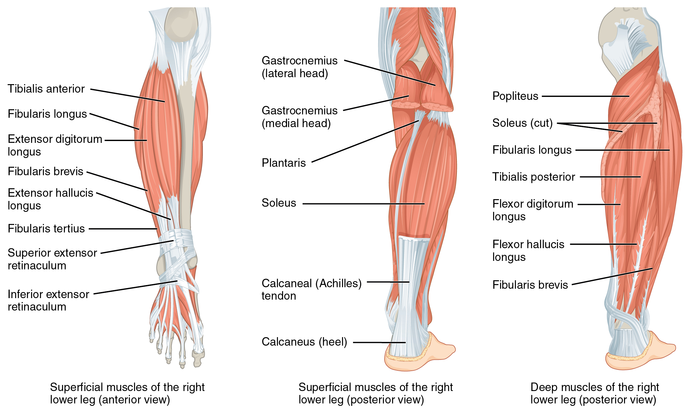
Tibialis Anterior Muscle
| Muscle | Origin | Insertion | Innervation | Action |
|---|---|---|---|---|
| Tibialis Anterior | Tibia (Upper 2/3 of lateral surface) IO membrane Superficial crural fascia |
Medial cuneiform (Medial & plantar surface) 1st Metatarsal (medial base) |
Deep Fibular n. L4 - L5 |
TCJ: DF STJ: Inversion |
Origin
Insertion
Functionally, the anterior tibialis inserts on the convex aspect of the medial arch7.
Nerve
Deep Fibular N. (L4, L5)6
Action
- Talocrural joint: Dorsiflexion
- Subtalar joint: inversion (supination)
The tibialis anterior works more as a supinator rather than an adductor7.
Observation
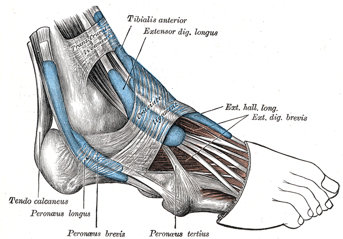
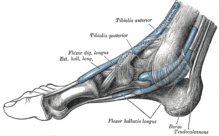
“The tendon of the tibialis anterior is visible at the level of the medial cuneiform and the base of the first metatarsal bone, especially if the foot is positioned in dorsiflexion and supination”8
Examination
MMT
- DF/inv
- “The tibialis anterior muscle produces the motion of dorsiflexion and inversion. The knee must remain flexed during the test to allow complete dorsiflexion. The patient’s foot is positioned in dorsiflexion and inversion. The leg is stabilized, and resistance is applied to the medial posterior aspect of the forefoot into plantar flexion and eversion”8
ROM
- PF + EV
Palpation
Pain Referral Pattern
Tibialis anterior pain can refer to the 1st toe or in the anterior leg over the muscle belly9.
Shin splints
Tibialis anterior dysfunction can lead to shin splints, more specifically anterior shin splints.
References
1.
Betts JG, Blaker W. Openstax Anatomy and Physiology. 2nd ed. OpenStax; 2022. https://openstax.org/details/books/anatomy-and-physiology-2e/?Book%20details
2.
Gray H. Anatomy of the Human Body. 20th ed. (Lewis WH, ed.). Lea & Febiger; 1918. https://www.bartleby.com/107/
3.
Donnelly JM, Simons DG, eds. Travell, Simons & Simons’ Myofascial Pain and Dysfunction: The Trigger Point Manual. Third edition. Wolters Kluwer Health; 2019.
4.
Neumann DA, Kelly ER, Kiefer CL, Martens K, Grosz CM. Kinesiology of the Musculoskeletal System: Foundations for Rehabilitation. 3rd ed. Elsevier; 2017.
5.
Weinstock D. NeuroKinetic Therapy: An Innovative Approach to Manual Muscle Testing. North Atlantic Books; 2010.
6.
Gilroy AM, MacPherson BR, Wikenheiser JC, Voll MM, Wesker K, Schünke M, eds. Atlas of Anatomy. 4th ed. Thieme; 2020.
7.
Jones B. B Project Foundations. b Project; 2025.
8.
Dutton M. Dutton’s Orthopaedic Examination, Evaluation, and Intervention. 5th ed. McGraw Hill Education; 2020.
9.
Myers HL, Devine WH, Fossum C, et al. Compendium Edition: Clinical Application of Counterstrain. Compendium ed. Osteopathic Press; 2012.
Citation
For attribution, please cite this work as:
Yomogida N, Kerstein C. Tibialis Anterior
Muscle. https://yomokerst.com/The
Archive/Anatomy/Skeletal Muscles/Lower limb muscles/Knee and Lower
Leg/Anterior compartment/tibialis_anterior.html