Spinal Cord
Pathway
The spinal cord exits the foramen magnum and extends to approximately the L1 vertebral level2.
Composition
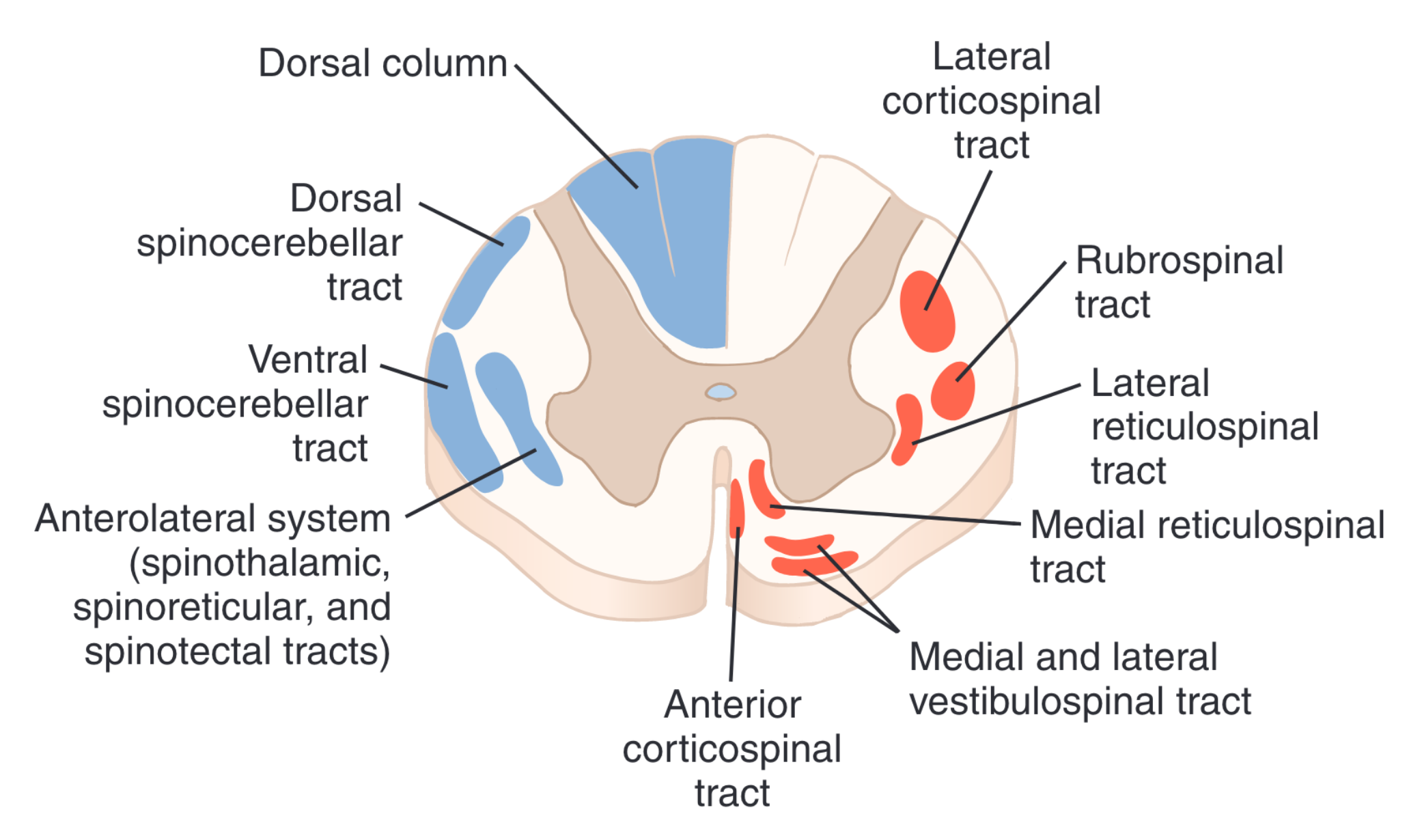
The spinal cord is made up of both white and gray matter.
White matter
- The white matter is solely composed of myelinated nerves
- The majority of the white matter consists of the ascending (sensory) tracts and descending (motor) tracts2.
- The rest of the white matter is composed of myelinated axons of interneurons.
Gray matter
The gray matter is found in the center of the spinal cord with a butterfly shaped horizontal cross section. The gray matter has 3 divisions:
- Ventral (Anterior) horn
- Intermediate Zone
- Dorsal (posterior) horn
The ventral (anterior) horn of the spinal cord contains the anterior motor neurons which
Motor Organization
Anterior motor neurons
Alpha Motor Neurons {#alpha-motor-neuron}
The α-motor neurons give rise to large type A α-motor nerve fibers (Aα) which have a diameter of ~14 µm3. These Aα-motor neurons branch multiple times after* entering the skeletal muscle and provide innervation to the large skeletal muscle fibers3. Since a single α-motor neuron gives rise to the large Aα-motor neurons which branch multiple times, the stimulation of a single α-motor neuron acn excite up to 100s of skeletal muscle fibers3.
The single α-motor neuron, its associated large Aα-motor neurons, and all of the skeletal muscle fibers make up a single motor unit3.
Gamma Motor neuron (γ)
Gamma (γ) motor neurons work alongside α-motor neurons to excite andcontract skeletal muscle fibers3.
γ-motor neurons are much smaller in size and there are 50% less neurons in the anterior horn compared to α-motor neurons3.
The γ-motor neurons give rise to even smaller Type-A γ-motor fibers which are ~5µmeters in diameter3. These fibers run to special skeletal muscle fibers known as “intrafusal fibers”3. These fibers can be smaller than the Aα-motor neurons since the intrafusal fibers are much smaller skeletal muscle fibers, compared to extrafusal fibers that the A&alpha-motor fibers innervate3.
These Aγ-motor neurons play a key role in controlling skeletal muscle tone3.
Interneurons
“Interneurons. Interneurons are present in all areas of the cord gray matter—in the dorsal horns, the anterior horns, and the intermediate areas between them, as shown in Figure 55-1. These cells are about 30 times as numerous as the anterior motor neurons. They are small and highly excitable, often exhibiting spontaneous activity and capable of firing as rapidly as 1500 times per second. They have many interconnections with one another, and many of them also synapse directly with the anterior motor neurons, as shown in Figure 55-1. The interconnections among the interneurons and anterior motor neurons are responsible for most of the integrative functions of the spinal cord that are discussed in the remainder of this chapter. Essentially all the different types of neuronal circuits described in Chapter 47 are found in the interneuron pool of cells of the spinal cord, including diverging, converging, repetitive-discharge, and other types of circuits. In this chapter, we examine many applications of these different circuits in the performance of specific reflex actions by the spinal cord. Only a few incoming sensory signals from the spinal nerves or signals from the brain terminate directly on the anterior motor neurons. Instead, almost all these signals are transmitted first through interneurons, where they are appropriately processed. Thus, in Figure 55-1, the corticospinal tract from the brain is shown to terminate almost entirely on spinal interneurons, where the signals from this tract are combined with signals from other spinal tracts or spinal nerves before finally converging on the anterior motor neurons to control muscle function.”3
Cross- Section
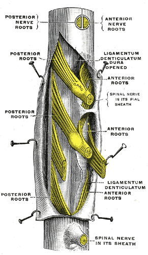
Spinal Nerve Roots
- 31 pairs of spinal nerves:
Cervical
Thoracic
Lumbar
Nerve root direction

One should take note that the nerve root direction changes as you descend the spinal cord. This is due to differing rates of growth of the spinal cord and vertebral column during development. Since the vertebral column elongates at a faster rate than the spinal cord, the spinal cord is essentially “drawn upward”2. In adults the spinal cord technically ends at L1 in the conus medullaris, but due to the elongation of the vertebral column, the nerve roots must travel inferiorly at an almost vertical direction2. This grouping of individual nerve roots travelling inferiorly creates the appearance of a “horse’s tail” and is referred to as the “Cauda Equina2.”
Due to the differing lengths of the spinal cord and vertebral column, more caudal injuries of the vertebral column will not directly correspond to the neurologic level of injury (NLI)2.