| Muscle | Origin | Insertion | Innervation | Action |
|---|---|---|---|---|
| Multifidus | ||||
| Rotatores breves | ||||
| Rotatores longi | ||||
| Semispinalis capitis | ||||
| Semispinalis cervicis | ||||
| Semispinalis thoracis |
Neck and Head MSK
Musculoskeletal Overview

Segments
The cervical spine can be divided into 3 segments:
- Upper
- Middle
- Lower
Upper consists of the first 2 cervical vertebrae: atlas (C1) and axis (C2).
The Middle segments refer to C3-C6 and technically begins at the inferior surface of axis (C2)5.
The Lower segment refers to the transition between the last cervical vertebrae (C7) and the first thoracic vertebrae (T1)5.
Movement
- Rotation
- Sidebend
- Flexion
- Extension
Rotation
Pure rotation
- atlanto-axial: Rotation + flexion
- Occipitoatlanto joint: Extension to offset the AA flexion5.
Joints
| Joint | Extension | Flexion | Sidebend | Rotation |
|---|---|---|---|---|
| Occipitoatlanto Joint | 10°2 15-20°5 |
5°2 10°5 |
~5° | Negligible |
| Atlanto-axial Joint | 10° | 5° | Negligible | 35-40° |
| C2-C7 | 55-60° | 35-40° | 30-35° | 30-35° |
| Craniocervical total | 75-80° | 45-50° | 35-40° | 65-75° |
| Joint | Extension | Flexion | Sidebend | Rotation |
|---|---|---|---|---|
| Occipitoatlanto Joint | 15-20°5 | 10°5 | ~8° | 12° |
| Atlanto-axial Joint | Negligible | 12° | ||
| C3-C7 | ° | ° | ° | ° |
| Cervical total | ° | ° | 45° | 80-90° |
Capsular pattern
In the cervical spine, when there is a capsular restriction here is the pattern of joint limitation:
- Sidebend and/or Rotation limited most/first
- Extension
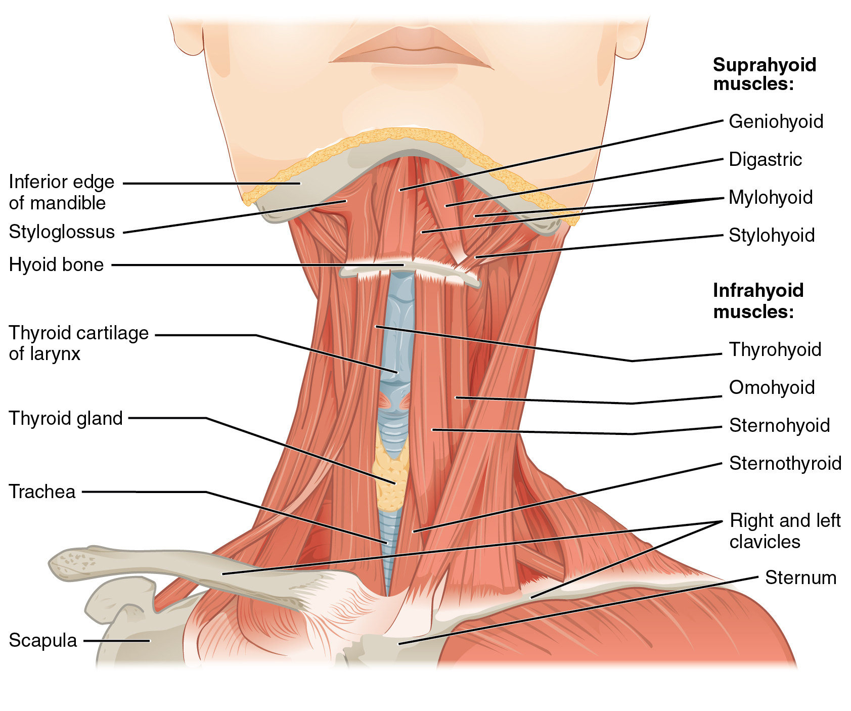
Muscles
The transversospinalis muscles run obliquely and medially from the transverse process of the inferior segment to the the spinous process of the superior segment5.
Unilateral contraction creates contralateral rotation5.
Bilateral contraction acts to cancel out rotation and create spine extension5. This extension accentuates the cervical lordosis created by the erector spinae5.
This muscle group functions in spinal stability, proprioception, posture, and stabilization of the vertebral column5.
The SCM can be seen as the “psoas of the neck” due to the fact it is commonly guarded or tight, but is rarely the cause of the overall dysfunction5.
Longissimus thoracis should be considered when viewing the neck since its uppermost fibers overlap medially with the iliocostalis cervicis5.
Anatomy
Intervertebral Disc (IVD)
Note
Named after vert above it6
The main dysfunction at the IVD is a herniation
Kinematics
Head Flexion
Osteokinematics: Flexion: 45-50 deg
Arthrokinematics:
Initial phase: begins in low c spine(C4-7) ,
Second phase: occurs initially in C0-2 then c2-3, C3-4
Flexion is described as an anterior osteokinematic rock (tilt) of the superior vertebra in the sag- ittal plane, a superoanterior glide of both superior facets of the zygapophyseal joints, and an anterior translation slide of the superior vertebra on the IVD
Uncovertebral joint: anterior spin6
Restriction: Anterior osteokinematic motion restricted by: PLL, interspinous, lig flavum, and extensor mm
Head Extension
Arthrokinematics:
- Posterior osteokinematic sagittal rock, an inferoposterior glide and approximation of the superior facets of the zygapophyseal joints, and a posterior translation of the vertebra on the disk.
- Uncovertebral joint undergoes a posterior arthrokinematic spin osteokinematic motion of extension is restricted by anterior prevertebral mm and ALL
Head Lateral SB
Osteokinematics: Side bend: 40 deg
Note
Sidebending: closer to 75 in supine because of hte muscles that limit SB with gravity: traps, scalenes, SCM (all are more lengthened in standing/sitting bc gravity pulling at insertions)(Harper 2023)
Arthrokinematics
- Superoanterior glide of contra superior facet posteroinferior glide of ipsi facet
- C/L translation of vertebra on disk
- Inferomedial glide of ipsi uncovertebral joint, superolateral glide of contral uncovertebral joint Limited by contra scalenes, intertransverse ligs, facet jt motions limited by joint capsule and translation limited by IVD
Head rotation
Rotation 70-90 deg Virtually all rotation in UPPER C/S occurs between Atlas & Axis
Arthrokinematics
Muscles: Global neck mm: SCM and semispinalis capitis and splenius captius Local neck: longus captius and colli, semispinalis cervicis, multifidus longus colli and post neck mm for a sleeve that supports spinal segments during mvmt
Tip
*same as sidebending motions
Coupled Motion
- C0-C2: Opposite
- C3-T3: Same
- T4-L5: opposite
Note
Coupled motion has been observed to change with age
Joints
OA Joint
OA joint Hypermobility is considered a diagnosis If range of rotation exceeds 8 degrees → stability testing necessary
AA Joint
Pure axial rotation 60% of total rotation in CS is here
Kinematics
Flexion
Greatest at lower C/S (C5-C6)
Least flexion at C2,3
Extension
Rotation
AA Joint performs pure axialrotation
Rotation and SB are coupled motions
Lateral flexion (SB)
Rotation and SB coupled motions
Coupled motion
Upper c spine: Sb and rotation — type 1 motion, OPPOSITE MOTION Regardless of posture Mid cervical: c3-c7 Same direction More type 1 motion: Changes in couple patterns
Clinical signifiance
- If c spine motion is limited w sb and rotation to same side
- 0 mid/lower c facet is suspected (restriction)
- Assess A/PROM
- Soft tissue
Posture
Cervical Lordosis
- C4/5 is midpoint of curve
- COG for skull is ant to foramen magnum
Dysfunction
Vestibular and Visual symptoms
- Upper c spine has more connections to vestib and visual systems than low c spine
- So upper c spine dysfunction can cause more balance/visual disturbances
Sensorimotor control
Tip
Wearing a hard collar for 5 days has been shown to lead to altered eye mvmt control, increased postural sway and disturbed head neck awareness in healthy peoplekristjanssonSensorimotorFunctionDizziness2009?
Examination
Screening
Warning signs of cervical region:
- Subjective
- Unexplained weight loss
- SB away from painful side that causes pain (if this is the only motion causing pain)
- Evidence of compromise of 2-3 spinal nerve roots
- Pain
- Gradual inc in pain
- Expansion of pain in terms of the regions involved
- Arm pain in pt younger than 35 or pt for more than 6 months
- ROM: Spasms w PROM
- Motor
- painful/weak resistive testing
- Limited scapular elevation
- T1 palsy (weakness / atrophy of the intrinsic mm of hand)
- Visual disturbances
- Hoarseness
- Horner syndrome–
- VBI and cervical myelopathy
ROM
- Upper C/S isolate rotation
- Cervical rotation Full Flexion
Special Tests
- VBI Test
- Sharp-Purser Test
- Alar Ligament Test
- Tectorial Membrane integrity
- Spurling’s Test
- TOS
- Allen’s Maneuver (should be performed before other TOS tests)
- CRLF Test (Lindgren’s test)
- Adson’s Test (TOS)
- ROOS Test
- Canadian C-Spine Rules
Provocative tests
- Disk herniation
- Vertebral end plate fracture
- Vertebral body fracture
Evaluation
- 1st Rib mobility
- Shoulder mobility
- Vertebra mobility
Evaluation inventory
Supine
- Spinous processes
- Alignment
- Depth
- A flat portion between segments is indicative of a tight/contracted muscle in that area bringing those segments together
- Suboccipital release
- If you cannot perform the release, loosening other things and returning to it would be a valid test-retest5.
- Sidebend mobility at each segment (compare R to L)
- Suboccipital opening (sidebend + rotation)
- Head pivoting on cervical spine
Supine with Flexed head
Bring the head into flexion and keep it there using your chest.
- Palpate cervical and head extensors
Manual Treatment
- Suboccipital release
- Work on each suboccipital individually
- PA Glides
- Upglides
- Downglides
- STM + Trap stretch
- STM + Levator scap stretch
- STM Scalenes
- STM + scalene stretch
- SCM STM
- ULTTs
- ULTT 1
- ULTT 2a
- ULTT 2b
- ULTT 3
Exercises
- DNF
- Shoulder Y, W, Ts
- Hemi Should Y,W, T, WER with cervical retraction
Cervical Radiculopathy
Wainner et al 2003 Cervical radiculoapthy diagnostic tests
- Positive ULTT1 (median nerve)
- Involved sign cervical rotation range of motion < 60 deg Positive distraction test
- Positive spurlings test
- 4/4 = 99 % specific, 30 x more likely to have cervical radiculopathy
- ¾ = 94% specific, 6 x more likely to have cervical radiculopathy
DDX
Reproduction of Pain with Cervical Distraction
- Spinal lig tear
- Annulus fibrosis (AF) Tear or inflammation
- Mm spasm
- Large disk herniation
- Dural irritability
Compression
- Arthritis
- Nerve root irritation
- Herniation
- Vert end plate fx
- vertebral body fx
Bearing down
- Reproduction of pain = C/s herniation
Pain
References
1.
Betts JG, Blaker W. Openstax Anatomy and Physiology. 2nd ed. OpenStax; 2022. https://openstax.org/details/books/anatomy-and-physiology-2e/?Book%20details
2.
Neumann DA, Kelly ER, Kiefer CL, Martens K, Grosz CM. Kinesiology of the Musculoskeletal System: Foundations for Rehabilitation. 3rd ed. Elsevier; 2017.
3.
Heick J, Lazaro RT. Goodman and Snyder’s Differential Diagnosis for Physical Therapists: Screening for Referral. 7th edition. Elsevier; 2023.
4.
Wise CH, ed. Orthopaedic Manual Physical Therapy: From Art to Evidence. F.A. Davis Company; 2015.
5.
Jones B. B Project Foundations. b Project; 2025.
6.
Dutton M. Dutton’s Orthopaedic Examination, Evaluation, and Intervention. 5th ed. McGraw Hill Education; 2020.
7.
Donnelly JM, Simons DG, eds. Travell, Simons & Simons’ Myofascial Pain and Dysfunction: The Trigger Point Manual. Third edition. Wolters Kluwer Health; 2019.
8.
Myers HL, Devine WH, Fossum C, et al. Compendium Edition: Clinical Application of Counterstrain. Compendium ed. Osteopathic Press; 2012.
Citation
For attribution, please cite this work as:
Yomogida N, Kerstein C. Neck and Head MSK. https://yomokerst.com/The
Archive/MSK/Regions/Spine/neck_MSK.html